January 2020
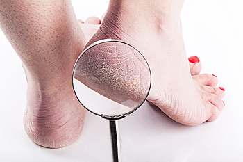
Solutions for Cracked Heels
Cracked heels may make you want to think twice about showing off your feet in warmer weather. However, cracked heels may be harmful to more than just the appearance of your feet. If deep fissures and cracks develop in your heels, they may make walking and standing painful for you. Additionally, these openings make way for germs to enter through your skin and cause infection.
There are several different causes of cracked heels. One of the most common reasons for this ailment is dry skin. This problem may make your keeps feel rough tight and itchy. Dry skin may be caused by cold air, extremely hot water, harsh soaps, and aging. Skin disorders such as eczema and psoriasis may eventually lead to dry skin. In some cases, complications may arise from cracked heels. Some of these complications are a loss of feeling in the heel, cellulitis, or a diabetic foot ulcer.
There are ways you can try to prevent getting cracked heels. One of the best ways to do so is to avoid wearing flip flops and sandals because these shoes increase your risk of drying out your feet. You should also avoid wearing shoes with a tall skinny heel, because these shoes cause your heel to expand sideways. At night, you should slather on a thick moisturizing cream on your feet and then cover them in socks to keep your feet moisturized overnight. Drinking water to stay hydrated is also a good way to ensure that your skin doesn’t become dry.
If you suffer from a severe case of cracked feet, you should make an appointment with your podiatrist to see what treatment methods are best for you.
Can Medical Conditions Cause Cracked Heels?
 A common cause of cracked heels often begins with dry skin. As this progresses, the skin on the outer edges of the heel may bleed, causing pain and discomfort. This can happen as a result of environment, genetics, and medical conditions that can include diabetes and thyroid concerns. It may become worse as daily activities are accomplished, and this may be a result of the weight the heel endures while standing and walking. Many patients have found relief when the feet are washed and dried thoroughly, followed by applying a good moisturizer. For mild cases of cracked heels, it may be beneficial to soak the feet in warm water to soften the skin. If you have the beginning symptoms of cracked heels, it is suggested that you consult with a podiatrist who can offer you proper treatment techniques.
Cracked heels are unsightly and can cause further damage to your shoes and feet. If you have any concerns, contact the podiatrists from Issaquah Foot & Ankle Specialists. Our doctors can provide the care you need to keep you pain-free and on your feet.
Cracked Heels
Cracked heels appear unappealing and can make it harder for you walk around in sandals. Aside from looking unpleasant, cracked heels can also tear stockings, socks, and wear out your shoes. There are several methods to help restore a cracked heel and prevent further damage.
How Do You Get Them?
Dry skin is the number one culprit in creating cracked heels. Many athletes, walkers, joggers, and even swimmers suffer from cracked heels. Age and skin oil production play a role to getting cracked heels as well.
Promote Healing
Over the counter medicines can help, especially for those that need instant relief or who suffer from chronic dry feet.
Wear Socks – Wearing socks with medicated creams helps lock in moisture.
Moisturizers – Applying both day and night will help alleviate dryness which causes cracking.
Pumice Stones – These exfoliate and remove dead skin, which allows for smoother moisturizer application and better absorption into the skin.
Change in Diet
Eating healthy with a well-balanced diet will give the skin a fresh and radiant look. Your body responds to the kinds of food you ingest. Omega-3 fatty acids and zinc supplements can also revitalize skin tissue.
Most importantly, seek professional help if unsure how to proceed in treating cracked heels. A podiatrist will help you with any questions or information needed.
If you have any questions, please feel free to contact one of our offices located in Issaquah, WA . We offer the newest diagnostic and treatment technologies for all your foot care needs.
Dr. Timothy Young's Postoperative Swelling Tips
After foot surgery, it’s not uncommon to have edema that can last from just 2 or 3 weeks, up until 2 or 3 months or even longer. It is always advantageous to try to reduce swelling when possible. There are multiple strategies for reducing swelling. Reducing swelling very often helps reduce postoperative discomfort and reduces the need for post procedure pain medications. Note postoperative swelling can be persistent. Once one or several techniques are done to reduce it, an effort has been made to maintain this because the swelling tends to want to keep coming back. This will happen until several weeks or longer have gone by and additional soft tissue and/or bone healing has occurred.
Outside physical therapy: We often wait for outside physical therapy until least 3 weeks postoperatively because this gives the incision to get a chance to have some initial healing to "seal up" the wound so to speak and to therefore minimize the risk of a postoperative infection.
Home treatments:
RICE: Rest Ice Compression and Elevation - this is always helpful. But our patients often forget.
Rest: Avoid going back to work or doing activities too soon. After initially being a couch potato for the first 3-5 days, some people just get too busy, and some people start to resume full weightbearing activities (too soon) with one's feet dependent or hanging down allowing gravity to pull more fluid down to the feet and ankles. This is often a time when initially you are feeling great, and then you start to overdo it and all of a sudden it starts hurting again and it may start swelling also. Remember, if you have to go back to work and your feet start to swell, you may have to compensate by really pampering yourself at home. Often it is helpful to go back to work for reduced hours during the first few weeks of returning to work. For example for major foot surgery, working just 4 hours per day 3 days a week the first week, than 4 hours per day 5 days a for several weeks, and then returning to work 6 hours 5 days a week, and finally a full 8 hour shift.
Ice: Whenever there is swelling, ice can be of some help. It's usually the most helpful during the first 48 hours of an injury but with surgery this can be more like the first 7 days.
Elevation: In particular, whenever possible if you're just sitting, try to keep your feet elevated above heart level.
Compression: An Ace wrap office compression. Sometimes it's helpful to do fairly intense compression with the Ace wrap leave it on for 20 minutes and then loosening the Ace wrap to more moderate pressure.
Remember to never remove the dressing including Ace wrap and last unit if the doctor is okay during the first 3-4 days. Remember, Ace wraps can lose some of their compression over time.
There are special forefoot compression sleeves that we can order or may be available through our clinic.
Contrast baths: After the first 1 to 2 weeks, there can be a benefit to doing contrast baths. Ask Dr. Nelson or Dr. Young before doing this. But, one technique is to have a very light sterile dressing on the foot, and then use a brand-new garbage bag and have the foot go into one top of warm water for 3-4 minutes at one top of his water for 1-2 minutes. This is a 5 minute cycling total, the 5 minutes cycled is repeated for 20-30 minutes.
Home interferential electrical stimulation (IF 4000 unit). These units are often available to check out from our clinic. These use a set of 4 gel electrodes that are placed per your doctors’ recommendations. Typically these are done for 20-30 minutes 1-2 times a day. Also typically icing is done during this treatment and/or immediately after this treatment. The units are usually used for 2-3 weeks duration. This helps reduce swelling and pain both.
Massage and ROM (range of motion): Gentle massage away from the surgical site can be very beneficial. One technique is for example, if bunion surgery was done then the side of the foot adjacent to the great toe is avoided. But the outside of the foot that may still have some swelling and/or edema is gently massage well the foot is elevated above heart level. This massage is worked from the toes back toward the midfoot toward the ankle and then up towards the leg. Most of the time is concentrating on the foot and ankle. The goal is to physically work some as swelling out by gently manually "moving" the fluid. After this can be very beneficial to place a compressive sleeve and/or Ace wrap over the foot to help avoid the swelling from immediately returning.ROM can be very helpful especially well the foot is elevated above heart level.
Supplements: It is always helpful to take supplements the help of bone healing such as calcium, magnesium, vitamin D 3 and vitamin K 2.
But in addition, it can help to take other supplements such as Omega essential fatty acids (fish oil), some people swear by tumeric and Arnica.
Rx Oral anti-inflammatories: Some anti-inflammatories may have properties that inhibit and/or slow bone healing. Taking Advil or Aleve once in a while should not be a problem, but taking it consistently or taking prescription anti-inflammatories can be a concern.
Hyperhidrosis of the Feet
Hyperhidrosis of the feet, also termed plantar hyperhidrosis, is characterized by excessive sweating of the feet that can be onset by any cause, such as exercise, fever, or anxiety. Most people suffering from hyperhidrosis of the feet also experience hyperhidrosis of the hands, or palmar hyperhidrosis. Approximately 1-2% of Americans suffer from this disorder.
Sweating is a healthy process utilized by the body in order to cool itself and maintain a proper internal temperature, which is controlled by the sympathetic nervous system. In individuals with hyperhidrosis, the sympathetic nervous system works in "overdrive", producing far more sweat than is actually needed.
Plantar hyperhidrosis is considered primary hyperhidrosis. Secondary hyperhidrosis refers to excessive sweating that occurs in an area other than the feet, hands, or armpits, and this indicates that is related to another medical condition, such as menopause, hyperthyroidism, or Parkinson's disease.
Symptoms of hyperhidrosis of the feet can include foot odor, athlete's foot, infections, and blisters. Because of the continual moisture, shoes and socks can rot which creates an additional foul odor and can ruin the material, requiring shoes and socks to be replaced frequently. In addition to the physical symptoms, emotional health is often affected as this disorder can be very embarrassing.
If left untreated, hyperhidrosis will usually persist throughout an individual's life. However, there are several treatment options available. A common first approach to treating hyperhidrosis of the feet is a topical ointment. Aluminum chloride, an ingredient found in antiperspirants, can be effective at treating hyperhidrosis if used in high concentration and applied to the foot daily. Some individuals can experience relief this way, while others encounter extreme irritation and are unable to use the product. Another procedure is the use of Botulinum Toxin A, commonly referred to as Botox. This is injected directly into the foot, and is effective at minimizing the sweat glands in the injected area. These injections must be repeated every 4 to 9 months.
If these treatments are ineffective, oral prescription medications may be taken in an effort to alleviate the symptoms. Again, some will experience relief while others do not. Going barefoot reportedly provides relief for most sufferers.
A final approach to combating hyperhidrosis of the feet is through surgery. Surgery has been less successful on patients with plantar hyperhidrosis than on those with palmar hyperhidrosis. It is only recommended when sweating is severe and other treatments have failed to work. This kind of surgery usually involves going into the central nervous system, and cutting nerves to stop the transmission of signals telling the foot to sweat.
Possible Causes of Plantar Hyperhidrosis
 Patients who have excessively sweaty feet may have a condition that is referred to as plantar hyperhidrosis. It may be caused by genetic factors, in addition to extreme emotional or physical stress. This condition may be treated by wearing inner soles that are absorbent, and the feet may feel better while using powders that can absorb the sweat. Additionally, it may help to change socks frequently, which may be beneficial in preventing an infection. If you are suffering from sweaty feet, it is advised that you seek the counsel of a podiatrist who can help you find the right treatment method.
If you are suffering from hyperhidrosis contact the podiatrists of Issaquah Foot & Ankle Specialists. Our doctors can provide the care you need to attend to all of your foot and ankle needs.
Hyperhidrosis of the Feet
Hyperhidrosis is a rare disorder that can cause people to have excessive sweating of their feet. This can usually occur all on its own without rigorous activity involved. People who suffer from hyperhidrosis may also experience sweaty palms.
Although it is said that sweating is a healthy process meant to cool down the body temperature and to maintain a proper internal temperature, hyperhidrosis may prove to be a huge hindrance on a person’s everyday life.
Plantar hyperhidrosis is considered to be the main form of hyperhidrosis. Secondary hyperhidrosis can refer to sweating that occurs in areas other than the feet or hands and armpits. Often this may be a sign of it being related to another medical condition such as menopause, hyperthyroidism and even Parkinson’s disease.
In order to alleviate this condition, it is important to see your doctor so that they may prescribe the necessary medications so that you can begin to live a normal life again. If this is left untreated, it is said that it will persist throughout an individual’s life.
A last resort approach would be surgery, but it is best to speak with your doctor to find out what may be the best treatment for you.
If you have any questions, please feel free to contact one of our offices located in Issaquah, WA . We offer the newest diagnostic and treatment technologies for all your foot care needs.
What Does a Partial Matricectomy Look Like?
Many of our patients especially are concerned about the cosmetic appearance of the toenail once the ingrown border is gone. Actually it is very difficult to even see this and be able to tell what side has been done permanently. Here is a photograph of a patient who had ingrown toenails removed (partial matricectomy procedures) from both the medial (inside) and lateral (outside borders) of the right great toenail and the medial or inside border of the left great toenail. See if you can tell the difference, most patients really can't tell the difference. Or they have to look very close to be able to tell the difference.
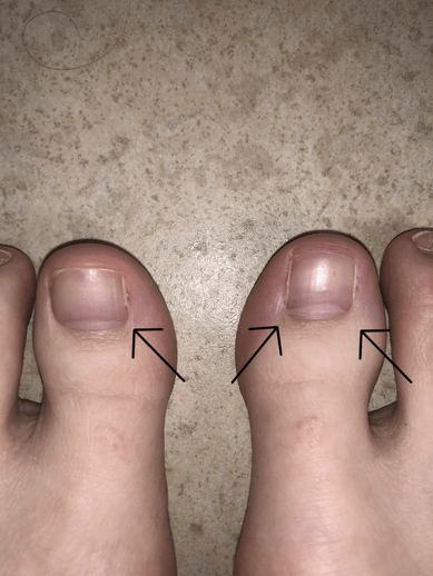
If you have questions about these treatments please let us know. We treat problems like this daily. Sometimes we see and treat 5 or more patients with this procedure on a given day. Give us a call at 425-391-8666 or make an appointment online today.
Neuromas - How To Treat Them
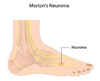
Morton's Neuroma (Intermetatarsal Neuroma)
Our Doctors have been using Alcohol injections for Morton's Neuroma for over 10 years with excellent success. Most patients have between 6 - 8 injections at weekly intervals. Occasionally a mini-series will be done after 12 months. For most patients, there is minimal discomfort after the injections and there is progressive relief from the Neuroma symptoms. We typically use between 20-30% dehydrated alcohol. Ultrasound guidance is critical for correct injection technique. Most frequently we treat the 3rd intermetatarsal space, and runner up is the 2 nd intermetatarsal space. This provides an excellent alternative to surgical treatment, and we also treat stump neuroma problems (where there is reoccurrence of the neuroma symptoms after prior foot surgery).
What Is a Neuroma?
A neuroma is a thickening of nerve tissue that may develop in various parts of the body. The most common neuroma in the foot is a Morton’s neuroma, which occurs between the third and fourth toes. It is sometimes referred to as an intermetatarsal neuroma. “Intermetatarsal” describes its location in the ball of the foot between the metatarsal bones. Neuromas may also occur in other locations in the foot.
The thickening, or enlargement, of the nerve that defines a neuroma is the result of compression and irritation of the nerve. This compression creates enlargement of the nerve, eventually leading to permanent nerve damage.
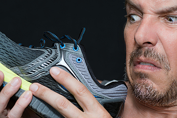
Causes Anything that causes compression or irritation of the nerve can lead to the development of a neuroma. One of the most common offenders is wearing shoes that have a tapered toe box, or high-heeled shoes that cause the toes to be forced into the toe box.
People with certain foot deformities – bunions, hammertoes, flatfeet, or more flexible feet – are at higher risk for developing a neuroma. Other potential causes are activities that involve repetitive irritation to the ball of the foot, such as running or court sports. An injury or other type of trauma to the area may also lead to a neuroma.
Symptoms If you have a Morton’s neuroma, you may have one or more of these symptoms where the nerve damage is occurring:
· Tingling, burning, or numbness
· Pain
· A feeling that something is inside the ball of the foot
· A feeling that there’s something in the shoe or a sock is bunched up
The progression of a Morton’s neuroma often follows this pattern:
· The symptoms begin gradually. At first they occur only occasionally, when wearing narrow-toed shoes or performing certain aggravating activities.
· The symptoms may go away temporarily by removing the shoe, massaging the foot, or by avoiding aggravating shoes or activities.
· Over time the symptoms progressively worsen and may persist for several days or weeks.
· The symptoms become more intense as the neuroma enlarges and the temporary changes in the nerve become permanent.
Diagnosis
To arrive at a diagnosis, the foot and ankle surgeon will obtain a thorough history of your symptoms and examine your foot. During the physical examination, the doctor attempts to reproduce your symptoms by manipulating your foot. Other tests or imaging studies may be performed.
The best time to see your foot and ankle surgeon is early in the development of symptoms. Early diagnosis of a Morton’s neuroma greatly lessens the need for more invasive treatments and may avoid surgery.
Non-surgical Treatment
In developing a treatment plan, your foot and ankle surgeon will first determine how long you’ve had the neuroma and evaluate its stage of development. Treatment approaches vary according to the severity of the problem.
· Icing. Placing an icepack on the affected area helps reduce swelling.
· Activity modifications. Activities that put repetitive pressure on the neuroma should be avoided until the condition improves.
· Medications. Oral nonsteroidal anti-inflammatory drugs (NSAIDs), such as ibuprofen, may be recommended to reduce pain and inflammation, or topical compounds.
· Over the Counter nerve medications. We highly recommend nutritional supplements that help with nerve inflammation.
· Bracing. Temporary offloading braces are helpful.
· Prescription custom orthotics devices. Custom molded prescription custom orthotics devices provided by your foot and ankle surgeon provide the support needed to reduce pressure and compression on the nerve.
· Injection therapy. Treatment may include injections of cortisone, local anesthetics or other agents. Usually the first line therapy is alcohol injections in combination with the prescription custom orthotics both together are extremely successful.
When Is Surgery Needed?
Surgery may be considered in patients who have not responded adequately to non-surgical treatments. Your foot and ankle surgeon will determine the approach that is best for your condition. The length of the recovery period will vary, depending on the procedure performed.
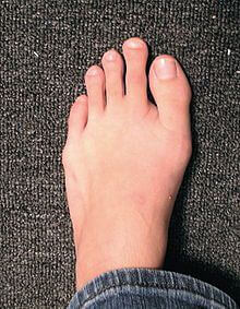
Bunions
A bunion is an enlargement of the base joint of the toe that connects to the foot, often formed from a bony growth or a patch of swollen tissues. It is caused by the inward shifting of the bones in the big toe, toward the other toes of the foot. This shift can cause a serious amount of pain and discomfort. The area around the big toe can become inflamed, red, and painful.
Bunions are most commonly formed in people who are already genetically predisposed to them or other kinds of bone displacements. Existing bunions can be worsened by wearing improperly fitting shoes. Trying to cram your feet into high heels or running or walking in a way that causes too much stress on the feet can exacerbate bunion development. High heels not only push the big toe inward, but shift one's body weight and center of gravity towards the edge of the feet and toes, expediting bone displacement.
A podiatrist knowledgeable in foot structure and biomechanics will be able to quickly diagnose bunions. Bunions must be distinguished from gout or arthritic conditions, so blood tests may be necessary. The podiatrist may order a radiological exam to provide an image of the bone structure. If the x-ray demonstrates an enlargement of the joint near the base of the toe and a shifting toward the smaller toes, this is indicative of a bunion.
Wearing wider shoes can reduce pressure on the bunion and minimize pain, and high heeled shoes should be eliminated for a period of time. This may be enough to eliminate the pain associated with bunions; however, if pain persists, anti-inflammatory drugs may be prescribed. Severe pain may require an injection of steroids near the bunion. Orthotics for shoes may be prescribed which, by altering the pressure on the foot, can be helpful in reducing pain. These do not correct the problem; but by eliminating the pain, they can provide relief.
For cases that do not respond to these methods of treatment, surgery can be done to reposition the toe. A surgeon may do this by taking out a section of bone or by rearranging the ligaments and tendons in the toe to help keep it properly aligned. It may be necessary even after surgery to wear more comfortable shoes that avoid placing pressure on the toe, as the big toe may move back to its former orientation toward the smaller toes.
What Is the Bump on the Side of My Big Toe?
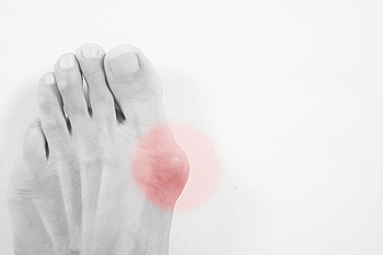 If you notice a bump on the side of your big toe, you may have what is referred to as a bunion. It may cause pain and discomfort, and it may be difficult to wear shoes that are typically worn on a weekly basis. Some of the symptoms that are generally associated with this condition can include calluses that form on top of the bunion, pain and swelling surrounding the affected area, and the skin may feel sore. Bunions have been known to be caused by wearing shoes that do not have ample room for the toes to move freely in. Additionally, there may be existing medical conditions such as gout or rheumatoid arthritis that can contribute to this condition. If you have developed a bunion, it is advised that you consult with a podiatrist who can offer you proper treatment options.
If you are suffering from bunions, contact the podiatrists of Issaquah Foot & Ankle Specialists. Our doctors can provide the care you need to keep you pain-free and on your feet.
What Is a Bunion?
A bunion is formed of swollen tissue or an enlargement of boney growth, usually located at the base joint of the toe that connects to the foot. The swelling occurs due to the bones in the big toe shifting inward, which impacts the other toes of the foot. This causes the area around the base of the big toe to become inflamed and painful.
Why Do Bunions Form?
Genetics – Susceptibility to bunions are often hereditary
Stress on the feet – Poorly fitted and uncomfortable footwear that places stress on feet, such as heels, can worsen existing bunions
How Are Bunions Diagnosed?
Podiatrists often perform two tests – blood tests and x-rays – when trying to diagnose bunions, especially in the early stages of development. Blood tests help determine if the foot pain is being caused by something else, such as arthritis, while x-rays provide a clear picture of your bone structure to your provider.
How Are Bunions Treated?
- Refrain from wearing heels or similar shoes that cause discomfort
- Select wider shoes that can provide more comfort and reduce pain
- Anti-inflammatory and pain management drugs
- Orthotics or foot inserts
- Surgery
If you have any questions, please feel free to contact one of our offices located in Issaquah, WA . We offer the newest diagnostic and treatment technologies for all your foot care needs.
Heel Pain Can Be Treated!

Do you suffer from heel pain when you get up in the morning? If so, you should seek professional help and have a proper diagnosis performed. Heel pain can be caused by several different foot-related conditions.
