General

  • Pain In The Ball Of The Foot
    Pain In The Ball Of The Foot 
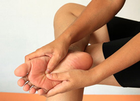
There are several causes of pain in the ball of the foot. The type of pain and its location help the doctor in determining the cause of the pain and helps to direct them in the best treatment for the patient.

    Calluses on the bottom of the foot can be painful. These calluses are caused by an abnormal alignment of the bones behind the toes called metatarsal bones. When there is an abnormal alignment of one or more of these bones, they will experience excessive weight and pressure. The skin thickens in response to this pressure forming the callus. Treatment consists of periodic trimming or shaving the callus, padding the shoes to remove the pressure, functional shoe orthotics, or surgery. Surgery should not be contemplated until the use of orthotics has been determined to have failed. The surgery consists of fracturing the involved metatarsal bone to realign it (See metatarsal surgery). Following the surgery, the patient should wear an orthotic to prevent the occurrence of new areas of callus. People with diabetes should not trim or shave these areas or use over the counter corn removers.

    Pain in the ball of the foot not associated with calluses can be a result of inflammation of a tendon into the toe, inflammation of the joint, or due to a pinched nerve called a Neuroma. A neuroma will also often cause a burning pain into one or more toes. Another cause of pain in the ball of the foot is arthritis. Arthritis will usually affect multiple joints in the ball of the foot. Common types of arthritis are rheumatoid arthritis, osteoarthritis and degenerative arthritis. Degenerative arthritis most commonly affects the big toe joint. The big toe joint is often stiff causing jamming (Hallux Limitus). Over time the jamming of the joint will cause an enlargement on the top of the joint.

    The most common cause of pain in the ball of the foot, in the area of the big toe joint, is sesamoiditis. Sesamoiditis is an inflammation of two small bones under the big toe joint. On occasion, a stress fracture can occur in one of these bones. Severe pain with sudden onset about the big toe joint may be gout.

  • Metatarsus Adductus

    Metatarsus adductus is a congenital deformity of the foot where there is increased curvature of the forefoot. This gives the foot the appearance of a "C" shape. This deviation of the metatarsals or visual effect of in-toeing is a deformity that occurs at the midfoot of the foot. The diagnosis of metatarsus adduction is relatively straightforward and is predominantly a clinical diagnosis. The exact cause of metatarsus adductus is not fully understood but is considered to be caused by intrauterine position and pressures. There may also be a genetic component to the deformity.

    Diagnosis

    The diagnosis of metatarsus adductus is made by physical examination and has certain characteristics:

    • the foot has an inward position as compared to the lower leg
    • the foot has a concave border medially and a convex border laterally
    • the metatarsus adductus foot may appear with a high arch
    • there is usually a separation of the big toe from the lesser toes

    Treatment

    Non Operative Treatment

    Most children with the metatarsus adductus deformity can be treated with conservative measures. Mild flexible cases can be treated with stretching and strengthening modalities, braces, orthoses, or straight last or reverse last shoe gear. In some cases serial casting may be necessary. This consists of applying plaster to the foot reducing the deformity. The cast is changed every 7 to 14 days. Casting is recommended until the deformity is reduced and then for half the amount of time again. In other words if it takes 6 weeks of casting to reduce the deformity then the infant should be cast for an additional three weeks. In those children who require casting those treated prior to the age of ambulation have more favorable results, but that doesn't mean that those with the deformity shouldn't be treated after ambulation begins. In cases where the deformity is resistant to conservative treatment or the deformity is rigid, surgical treatment may be considered.

    Operative

    Operative treatment is reserved for deformities that have been neglected or have not responded to non-operative treatments. If surgery is required there are several different procedures depending on the age of the patient and the magnitude of the deformity. In less severe cases, soft tissue releases or tendon transfers can correct the deformity. While in more severe cases bony cuts and repositioning is the best treatment option. Following any type of surgical correction braces, orthoses or orthopedic shoes may be required.

  • Lumps And Bumps On The Outside Of The Foot

    There are a few causes of lumps, bumps or prominence on the outside of the foot. Some of these are just normal anatomy; others are due to abnormal processes. Starting from the fifth toe and working back to the heel:

    A common prominence on the fifth toe is due to the formation of a hammertoe. A hammertoe is a structural deformity of the fifth toe causing the joint in the toe to become propionate. Pressure from shoes will cause a thickening of the skin. On occasion, this can become very painful making it difficult to wear a closed shoe. Treatment consists of trimming the thick skin or padding the area to reduce the pressure from the shoe. At times surgical strengthening of the toe is necessary (See hammertoe surgery).

    Just behind the fifth toe, at the joint where the toe connects to the foot, a prominence can develop. This is called a Tailors bunion. It is caused by the abnormal function of the foot and can be slowly progressive. Some people naturally have an enlargement in this area. Shoe pressure can make the area painful and at times cause a bursa to form. A bursa is a sack of inflamed tissue that occurs over a bony prominence in response to excessive pressure or friction. The bursa is spongy to the touch and can be very painful. The bursa can be treated with cortisone injections to reduce the swelling and inflammation. Padding the area is also useful to reduce the pressure from the shoes. Selecting the correct shoe can also be helpful (See selecting the appropriate shoe). Surgical correction of the tailor's bunion is a common procedure to reduce the prominence and the pain associated with it (See Tailors bunion surgery). A rare occurrence of gout can also occur in this area. This condition usually presents with a sudden onset of pain and swelling in the area.

    Further down, along the side of the foot another bony prominence can observed. This area is the base of the long bone (metatarsal bone) behind the fifth toe called the styloid process of the fifth metatarsal. In children this area has the growth plate to the bone. Injury to the area can cause the area to become enlarged. X-rays will show an open growth plate until the age of 12 - 14. If there has been an injury to this area in a child with an open growth plate, caution must be taken because of the possibility of injury to the growth plate. A very powerful tendon also attaches into this area of the bone. In the event of an ankle sprain this area can become injured as a result of the tendon attempting to prevent the foot from twisting. Pain to the touch at this site indicates either tendon or bone injury, and an x-ray should be taken. A fracture can occur in this area, and if it is displaced or out of alignment then surgery may be required to repair the bone. If the fracture is in good alignment or if the area is just sprained, then a below the knee cast should be applied. It may take six weeks for this injury to heal.

    Some people have a natural enlargement of this area. When painful, padding the area to reduce shoe pressure is useful and selecting the correct shoe is also helpful. Rare instances of gout can also occur in this area.

    Just behind the area of the styloid and a bit over to the top of the foot there is a spongy, soft area of the foot. This area represents the extensor digatorum muscle. This is the only muscle present on the top of the foot. In some people it can be quite large. Fatty deposits can also occur about the muscle making the area appear larger. On occasion, a ganglion may form. In this instance, the area may enlarge and then shirk and then enlarge again. A MRI is the most useful test to determine if a ganglion is present. If the area is not painful, no treatment is required. If the area is painful and a ganglion has been identified, then treatment might consist of draining the fluid from the ganglion with a needle or surgically removing the ganglion.

    Small, soft lumps can be observed around the fat pad of the heel when a person stands. These small, soft bumps are usually found in groups. They are called Piezogenic papules and represent herniations of fat from the heel fat pad. They are rarely painful and no treatment is necessary. Removal of the pziogenic papules should be discouraged. There is no adequate treatment for their removal, and surgery to the area will only make the condition worse.

    Another lump seen on the side of the heel is called an inclusion cyst. This lump appears as an elevated, soft mass which is often callused on is surface. These are caused by minor puncture wounds or from prolonged pressure or friction to the area by shoe gear. Surgical excision is often recommended. Padding the area to reduce friction may be of some temporary benefit in painful situations.

    Other less common lumps and bumps that can appear on the outside of the foot are lipomas, fibromas, and giant cell tumors. These are non-cancerous tumors that tend to slowly enlarge and have a spongy like consistency. They only present a medical problem if they interfere with the function of a tendon, press excessively on a nearby nerve or cause pain secondary to shoe pressure. Malignant tumors of the foot are exceedingly rare.

  • Lumps And Bumps On The Inside Of The Foot

    There are many different causes of lumps and bumps on the inside of the foot. Starting from the big toe and working back to the heel:

    A swelling along the margin of the big toenail is likely to be due to the formation of an ingrown toenail. At times this swelling can produce open, weeping flesh. Treatment consists of removing the ingrown toenail (See treatment for ingrown toenails).

    A swelling often associated with a hard, callused area may form on the side of the big toe. This is due to an enlargement of the bone in this area. It is usually associated with a deviation at the joint in the big toe. Treatment consists of using a functional foot orthotic to redirect the pressure from the area. Surgical removal of the underlying bony prominence or surgical straightening of the toe may be recommended. People who have diabetes must watch this area because over time the excessive pressure to the area from walking can cause the skin beneath the callused area to break down forming an ulceration that can become infected. Left untreated, the bone can become infected and in severe cases amputation of the toe may be necessary.

    Another area on the big toe, which can demonstrate a lump or bump, is the top outside portion of the toe at the level of the joint in the toe. This is usually seen in association with a contracture of the big toe making the joint more prominent. This prominence can become irritated due to shoe pressure. The condition is called a Hallux hammertoe. The word Hallux is the medical term for the big toe. If the deformity is flexible and the toe can be manually straightened, the condition can be corrected by a simple surgery that releases the tendon in the bottom of the toe. If the condition is rigid and the toe cannot be manually straightened, then a fusion of the joint may be necessary (See surgical correction of hallux hammertoe). Treatment should also include determining what caused the condition. Quite often it is due to some underlying functional problem in the foot and treatment of this condition with a functional foot orthotic should be considered. Other causes of a hallux hammertoe are previous surgery in the area, nerve damage or a neurological condition.

    A painful lump may also be found on the bottom of the big toe. This condition often presents as a hard callused area. Sometimes a hard lump may be felt beneath the callused area. The most common cause of this condition is a limitation of movement at the big toe joint while walking. If there is limitation of movement at this joint, then the big toe cannot bend upward as the heel comes off the ground while walking. As a consequence, there is excessive force placed on the bottom of the big toe and an enlargement of the bone in this area will occur secondary to the pressure. Another cause is the presence of an extra bone or piece of cartilage in the area. However, it is the limitation of movement in the big toe joint that causes the area to become enlarged and painful. People who have diabetes must watch these areas closely. Over time, the excessive pressure can cause this area to break down and ulcerate. As in the previous condition this can lead to infection and possible amputation. One way to check to see if there is a limitation of movement of the big toe joint is to perform the following test. While standing on a flat surface have another person try to bend the big toe upward. The joint that must move is the joint where the toe attaches to the foot. There is a joint in the middle of the big toe and this is not the one you are checking. If the big toe joint cannot be bent upward then a limitation of motion exists. Testing the movement at the joint without putting weight on the foot will give a false impression as to the available movement at the joint while you are walking. The limitation of movement of the big toe while walking can, over time create an arthritic condition in the joint. A bony mass may then form on the top of this joint as a result of jamming in the joint. This condition is called hallux limitus or hallux rigidus. Treatment for the painful lesion in the bottom of the big toe joint consists of using functional foot orthosis to correct the functional limitation of the big toe joint motion or may consist of surgical correction of the hallux limitus. Rarely is surgery to remove only the lesion on the bottom of the toe alone successful, because the cause of the initial problem still exists.

    Yet another area of bony prominence about the big toe joint is on the outside of the joint. The most common cause of this condition is called hallux abductovalgus or bunion. This is a common condition associated with deviation of the big toe towards the second toe. Treatment consists of padding the area to reduce shoe pressure, bigger shoes, orthotics and surgery (See surgical correction of bunion deformity). A sudden onset of pain in the area with or without a bunion may be due to gout. Gout is a metabolic condition that results in an elevation of the uric acid in the blood stream. Once the uric acid level reaches a certain point it will crystallize and leave the blood stream and deposit itself in a joint or other soft tissue location. Gout rarely occurs in women until after menopause or if they have had a hysterectomy. A sudden onset of pain in this area in women is usually associated with the formation of a bursa secondary to wearing a tight shoe. Treatment consists of wearing looser fitting shoes and taking an oral anti-inflammatory medication. Occasionally, a cortisone injection may be advised.

    Moving back towards the heel, another area of a common lump or bump is just in front of the weight-bearing portion of the heel. This prominence is usually a hard bony mass that protrudes outward. It is due to a bony enlargement of the Navicular bone. Some people have a natural enlargement in this area while other people have an extra bone in this area. This condition is often referred to as Os Navicularis. This condition is often associated with a flat foot deformity. A very powerful tendon from a muscle in the leg called the posterior tibial tendon partially attaches into this area. If the navicular bone is naturally enlarged or there is a second bone present then the tendon dose not function properly and cannot support the arch of the foot adequately. Over time, this will cause a weakening of the tendon causing an entire collapse of the arch of the foot. This condition called Posterior Tibial Tendon Dysfunction is a very serious condition that should be treated promptly and aggressively. The bony prominence of the navicular bone may cause secondary pressure in shoes resulting in pain. Treatment of the prominence of the navicular bone consists of padding to reduce shoe pressure, functional foot orthosis to treat the associated flat foot deformity and the possible onset of posterior tibial tendon dysfunction, or surgery. Following any surgical intervention in this area, the patient should be placed in a functional foot orthosis. The surgical removal of the bony prominence does not correct a flat foot or prevent the occurrence of posterior tibial tendon dysfunction.

  • Leg Length And Deformity Correction - Ilizarov Tec

    Professor Gavriil Abramovich Ilizarov graduated from medical school in the Soviet Union in 1943, near the end of World War II. After graduation, he was assigned to practice in Kurgan, a small town in western Siberia. He was the only physician within hundreds of miles and had little in the way of supplies and medicine. Faced with numerous cases of bone deformities and trauma victims due to the war, Professor Ilizarov used the equipment at hand to treat his patients. Through trial and error, with handmade equipment, this self-taught orthopedic surgeon created the magical combination that would cause the bones to grow again, similar to the adolescent growth state. Thus the Ilizarov technique was created.

    For almost 45 years, Ilizarov and his co-workers have been perfecting their apparatus and helping individuals with severe orthopedic abnormalities. Reports of dwarfs made taller, and birth defects corrected were first observed in Italy, and then presented in the United States where Professor Ilizarov's technique met initial skepticism.

    Today, Professor Ilizarov's methods are an acceptable means of correction of severe orthopedic deformities. Limb correction is a gradual process, which lengthens and straightens bone and soft tissue so a limb can function as normally as possible. There are many reasons why a person may be a candidate for limb correction. For many patients, the procedure is used to straighten or lengthen a bone. Others have a need because of a trauma, which has caused highly fragmented breaks, bone loss, or to encourage a bone which isn't healing.

    How the Procedure Works

    This method takes advantage of the body's remarkable ability to grow new bone tissue. It involves the surgical application of a circular metal frame called the Ilizarov fixator. Fixator rings are attached to each segment of the original bone through pins and wires, which hold each bone segment in place while new tissue is growing and maturing.

    There are two main phases to the correction process: correction/lengthening and consolidation. The lengthening phase is the time needed to gradually achieve the desired correction/length of the limb. The consolidation phase is needed for the new bone tissue to harden and mature. Each individual's body is different, but the total time of wearing the fixator is typically 4-12 months.

    Lengthening refers to the period of time it takes to "grow the bone." The lengthening phase begins after the surgeon cuts the bone and attaches the fixator. During this time the patient will be working with the physician and the physical therapist to make gradual adjustments to the fixator which increase the gap between the bone segments, adding "length" to the total limb. Over a period of months, new bone tissue will grow in the gap, ultimately hardening the area between the segment of the original bone.

    When the physician is satisfied with the length and position of the new bone, the consolidation phase begins. During this phase, the bone tissue matures and becomes solid. The patient still wears the fixator, but no adjustments are made. The consolidation phase is the longest part of the Ilizarov process. It takes twice as long for bone to harden as to does to lengthen it, so the consolidation phase typically doubles the time spent in the lengthening phase.

    Post Operative Course

    During the lengthening phase, the patient will be seen by the physician every few weeks to monitor the progression of correction of the deformity. On each visit, X-rays are taken to monitor the progress of the lengthening and to assess the quality of the new bone. Office visits may range from thirty minutes to two hours, depending on what work needs to be done.

    If a limb lengthening procedure is being performed, the patient will be required to assist in the process by making fixator adjustments at home. This is usually done every six hours. The physician will make sure that the patient is fully trained and understands how to make adjustments if need.

    At times, some patients experience some failure of the mechanics of the fixator. The patient may have broken wires, bent rods, or be unable to turn one of the "clickers". Through proper education of the physician and the patient, these problems can be solved without permanent sequela.


    Other Uses of the Ilizarov Technique

    Limb lengthening are not the only procedures that can be used with the Ilizarov frame. Ankle fusions, triple arthrodesis, midfoot fusions, etc. can be performed without prolonged cast immobilization and non-weight bearing. In most cases, partial to full weight bearing is suggested to encourage functional capabilities and healing.

    Charcot reconstruction of the ankle or rearfoot is a prime example in which a fusion procedure can be performed with the patient full weight bearing on the involved extremity almost immediately. This diminishes the stress that is placed on the opposite extremity, which is prone to trauma due to increased stress of the uninvolved side.

    Conclusion

    The patient attitude throughout the Ilizarov process is very important to its success. The patient will have to find ways to cope with discomfort, pain, and frustration, plus the natural ups and downs of emotions during this long process. Knowledge of the process prior to surgery ensures proper patient compliance and minimal pain. Each individual experiences pain differently, so it is difficult to talk about norms when performing this type of surgery.

  • Kohler's Disease

    Kohler's Disease is a spontaneous loss of blood supply to a bone in the foot called the Navicular bone. Dr. Kohler described it in 1908. The spontaneous loss of blood supply to a bone is called osteochondrosis. In later years Dr. Kohler was also associated with another osteochondrosis of the foot known as Freiberg's disease. Some texts refer to this condition as Kohler's II.

    Clinical features and Treatment

    Clinically, the presentation of Kohler's disease may be an incidental x-ray finding. Often, however, localized pain or a painful gait is noted. Occasionally mild swelling is seen. It is seen most commonly in males and most cases only affect one foot. Biopsy of the bone to make the diagnosis is not necessary.

    Complete recovery is almost always the rule; therefore, treatment efforts should be conservative. Cast immobilization provides satisfactory results. Reduced activities and foot orthotics (inserts for the shoes) have also proved effective. Most cases respond within 8 months. Follow-up studies after 30 years have shown no residual degenerative changes in spite of severe fragmentation and flattening of the navicular bone. It is interesting to note that Kohler's disease has been reported to be associated with "Tarsal Coalition" in this area. So it is important that proper follow-up be done.

    This article was adapted from the text book Foot and Ankle Disorders in Children edited by Dr. Steven DeValentine.

  • Infections, Diabetic Infections, Cellulitis

    Infection can be caused by a number of different agents. Athlete's foot is an infection of the skin caused by a fungus. Warts are caused by a viral infection of the skin. When most people think about infections they are thinking about infection caused by bacteria. There are numerous types of bacterial infection. Infection generally requires a break in the skin from a cut, abrasion, puncture wound or ulceration. The most common infection is caused two bacteria, Staphylococcus and Streptococcus. Both of these infections cause progressive tissue damage of varying degrees. People with diabetes can develop infections by several bacteria at the same time.

    Infection that occurs in the skin is called cellulitis. Deep infections that develop puss pockets are called abscesses. The most common bacterium that causes cellulitis is Streptococcus. These infections can become very serious and even life threatening. Cellulitis is characterized by spreading redness in the area with an increase in the temperature of the skin, often accompanied by fever and chills. People who suffer from venous stasis, chronic swelling in the legs, are prone to these infections. Cellulitis is also commonly seen in associated with athlete foot conditions. The athlete's foot causes small breaks in the skin, which can become infected, by the Streptococcus bacteria. Soft corns, particularly between the fourth and fifth toes can also become infected and cause cellulitis and or an abscess. Puncture wounds are very likely to become infected. This can result in a very dangerous deep abscess that can also infect the bone. A doctor should evaluate all deep puncture wounds as soon as possible. Simply cleaning the outside of the puncture wound is not enough to prevent infection. Oral antibiotics should be prescribed and the wound watched carefully. If there is any sign of infection, surgical cleaning of the wound should be preformed.

    People with diabetes are at particular risk of infection. In fact, people with diabetes spend more time in the hospital for foot infections than for any other reason. Corns and calluses on the feet of people with diabetes can break down and allow bacterial invasion of the tissue. In people with long standing open ulceration the underlying bone can become infected. Bone infections, called osteomyelitis, generally require surgery to remove the infected bone. These infections are very difficult to cure with oral or intra-venous antibiotics without also removing the infected bone. The presence of bone infection can be diagnosed with special tests such as bone scans, CT scans and MRI. These test are not 100% accurate however and the experience of the treating doctor becomes very important in making the correct determination as to the presence of bone infection.

    Gout, which is caused by an elevated blood uric acid level, can be confused with infection. This condition can cause sudden onset of pain, redness, and swelling of a joint or in the soft tissues. It is important to rule out an infection, particularly if it is in a joint. Rare instances of infection can occur without evidence of a break in the skin. The infection is carried to the site by the blood stream. Close examination of the patient may reveal an abscessed tooth that likely is the source of the infection. In other instances no source of the infection can be found.

    People who have joint replacements, heart valve replacements or who have a pacemaker must be very cautious regarding infections. They are at risk of an infection spreading from another site to the area of their joint replacement, heart valve replacement or pacemaker. Also, people who have mitral valve prolapsed must be cautious if they have infections because the infection can spread to the heart.

  • Glossary Of Podiatry Terms

    A
    abductor (muscle)
    achillobursitis
    aclasis – pathological continuity of structure
    acrodermatitis enteropathica
    acrokeratosis paraneoplastica – Bazex syndrome
    adductor (muscle)
    adiposis dolorosa – Dercum disease
    aDWF – ankle Doppler wave form
    AFO – ankle-foot orthosis
    alligator skin – ichthyosis congenita
    allylamines – e.g. Naftin (naftifine)
    angle, DMMA – distal metatarsal articular, Engel, Fowler-Philip, Kite, talo-first, Taygar
    ankylosis
    anserinoplasty
    antalgic gait
    AOFS hindfoot ankle score
    apophysitis
    aPVR – ankle pulse volume recording
    atavistic tarsometatarsal joint
    athlete’s foot – tinea pedis, ringworm of the feet
    atavism

    PROPER NOUNS:
    Abrikosoff tumor
    Alizarin sweat test
    Allen maneuver
    Apelqvist grades 1 through 4
    Ashurst-Bromer classification of ankle fracture
    Atasoy flap for nail injury repair
    Austin – bunionectomy, screw, wire

    B
    ball-and-ring device
    basal cell nevus syndrome
    basic fuchsin (in fungicides)
    BB – blunted and bowed
    bipivotal hinge
    bivalve of cast
    BK amputation/case – below-the-knee
    blebs
    bowing of bone
    bowleg; bowlegged – genu valgum
    brachymetatarsia
    bubble patch
    bunion (see proper names in second alpha section)
    bunionectomy
    bunionette
    bursitis – inflammation of the bursa of a joint
    bursotomy
    butenafine hydrochloride – Mentax (antifungal)

    PROPER NOUNS:
    Banks-Laufman incision
    Bannon-Klein implant
    Bardenheuer bifurcation procedure
    Bart-Phumphery syndrome
    Bazex syndrome – acrokeratosis paraneoplastica
    Beau lines
    Bechtol arthroplasty
    Bednar tumor
    Berman-Gartland procedure
    Betadine scrub
    Bier amputation
    Blair arthrodesis
    Blajwas-Schwartz-Marcinko irrigation/drainage system
    Bledsoe cast brace
    Blex contractile force curve
    Blount, brace, disease, staples
    Blucher opening
    Bodsky ischemia classification
    Boots: Acme, Denis Browne, Dingo, Gibney, 9-West, Red Dog, SACH, Sorel, Stride Rite,
    Technica, TheraBoot, Unna Gelcast/gelatin, Unna paste, Vasque
    Brace, dropfoot, (AFO), ischial weightbearing, Knight, Lyman-Smith, Milwaukee,
    Stabheilizer (for footdrop), Taylor, toe drop, weightbearing
    Brewster triple arthrodesis
    Brockman incision
    Brostrom procedure
    Bunion – hallux abductovalgus, last, tailor’s,
    Bunionectomy:
    Akin
    Austin
    Joplin
    Keller
    Kreuscher
    Lapidus
    Logriscino
    McBride
    Mitchell
    Reverdin
    scarf osteotomy
    Silver
    Stone
    tricorrectional
    Wu
    Z
    Brunnstrom stage I-V
    Bunnell, anvil, drill, needle, operation, probe
    Burns-Haney incision
    Burow triangle

    C
    calcaneal – pertaining to the calcaneus bone
    calcaneitis
    calcaneoapohysitis
    calcaneoastragaloid
    calcaneocavus
    calcaneocuboid
    calcanodynia
    calcaneofibular
    calcaneonavicular
    calcaneoplantar
    calcaneoscaphoid
    calcaneotibial
    calcaneovalgocavus
    calcaneus bone
    calcar, femorale, pedis
    calcarine
    calf-corset weightbearing
    calf-lacing, double-upright brace
    callosity – pertaining to a callus
    callus; calluses
    capillary refill (time)
    capsulitis
    cast brace/boot
    castaway orthotic
    CCE – clubbing, cyanosis, edema
    cellulitis
    cetylpyridinium Cl
    cheiropodalgia
    chemical matricectomy
    chevron osteotomy
    chloroxylenol
    chondroma – tumor of the chondral tissue
    clam, clamshell walker
    claudication
    clavus, clavi: corn, heloma, duras, hystericus, interdigital, mollis, secalinus
    clawfoot
    clawtoe(s)
    cleft foot
    clonus; ankle, toe
    clubfoot – see talipes
    coalition
    cookie; metatarsal c.
    coralline hydroxyapatite
    corn – clavus, hard, interdigital, kissing, soft
    counter of shoe
    coax, valga/varum
    crescentic osteotomy
    CRPS – chronic regional pain syndrome

    PROPER NOUNS:
    Calandruccio fixation device
    Cam Walker walking brace
    Can-Am brace
    Carville sandal
    Cam Walker CBWO screw
    Chambers osteotomy
    Chang-Miltner incision
    Charcot, arthrosis, foot, joint, restraint, procedure disease
    C. restraint orthosis walker
    Charcot-Marie-Tooth disease – CMT
    Charnley, arthrodesis, fixator
    Charnley-Mueller; arthroplasty/prosthesis
    Chopart amputation
    Chrisman-Snook ankle reconstruction
    Cincinnati incision
    Clayton procedure
    Clearz – a fungicide
    Coban dressing
    Cobbies shoewear
    Codman triangle
    Cole osteotomy for midfoot deformity
    Coleman block test
    Colonna-Ralston incision
    Comf-Orthotic insoles
    Converse athletic shoewear
    Comed footgear
    Compound W
    Cook walking brace
    Corfan shoe
    Cotrel-Dubousset brace
    Count’R-Force arch brace
    Couvelaire incision
    Crego elevator
    CROW brace
    CRPS – chronic regional pain syndrome
    Cryo Cuff
    Curtain incision
    Cutter implant
    Cymeline

    D
    danglefoot, aka dropfoot
    DCCT – Diabetes Control and Complications Trial
    decubitus ulcer
    dendritic synovitis
    desquamates
    DFO – dorsiflexion osteotomy
    dial-in cut osteotomy
    diabetic toe(s)-(foot or feet) – see ULCER
    diaphysis
    DIP joint; DIPJ – distal interphalangeal joint
    disk disease
    DMMA – distal metatarsal articular angle
    dorsomedial
    duckfooted – flatfooted
    duckwalk
    duck waddle

    PROPER NOUNS:
    Danis-Weber classification of ankle injury
    DarcoGel ankle brace
    Darier sign
    De Guglielmo disease – erythemic myelosis
    De Morgan spots
    Dejerine-Sottas disease
    Denis Browne, bar, boot, splint, shoe
    Dercum disease
    DesignLine orthotic
    Diab-A-Thotics
    Di Guglielmo disease – erythemic myelosis
    Dias-Tachdjian classification of physeal injury
    Dickinson approach
    Diebold-Bejjani osteotomy
    Di Guglielmo disease
    Donjoy RocketSoc ankle brace
    Downey-McGlamry procedure
    Dressflex orthotic
    Dr. Scholl’s foot products
    Dubreuilh melanosis circumscripta
    Dunn triple arthrodesis
    DuoFilm products
    Dupuytren, contracture, exostosis
    DuVries incision/repair
    Dwyer osteotomy
    Dynafo ankle-foot orthosis (or D. AFO)
    Dynasplint orthotics
    DynaStep

    E
    epiphysis, epiphyseal

    PROPER NOUNS:
    Eastersohn osteotomy for tailor’s bunion
    Easy Spirit shoewear
    Eaton-Lambert syndrome
    Eckert-Davis classification of peroneal tendon subluxation
    Eichenholtz stage
    Elastomull
    Elmslie procedure
    Engel angle
    Esmarch bandage/tourniquet
    Essex-Lopresti fracture
    Etonic shoes
    Evans calcaneal osteotomy
    Evazote

    F
    flaccid
    flatfoot
    flexion/extension
    fractures – (see list in Surgical Compendium)
    fungus

    PROPER NOUNS:
    Famolare shoes
    Farber disease
    Felty syndrome
    Flex Foot products
    Floam cushion
    Florsheim shoes
    Footmaxx orthotics
    Foot Medic products
    Forestier disease
    Fosnaugh nail biopsy
    Fowler-Philip angle
    Freer elevation
    Freiberg infraction (not infarction)
    Friedreich
    Forst H-block
    Fungus products: Fungatin, Fungi Clear, Fungi-Nail – sorcinol, salicylic acid

    G
    GAIT, antalgic, ataxic, gegenhalten, halting, spastic, scissor, three-point, Trendelenburg, two-point
    ganglion cyst
    gastrocnemius
    gastrocsoleus
    GENU – knee, recurvatum, varum – bowleg, valgus/valgum – knock knee
    gout – podagra; tophaceous. gouty arthropathy
    gun-barrel sign
    guttate keratoses

    PROPER NOUNS:
    Ganley splint
    Gowers sign

    H
    hammertoe(s)
    hamstring muscle/tendon
    HALLUX, dolorosa, flexus, limitus (limitans), malleus, rigidus, valgus, interphalangeus –HBO – hyperbaric oxygen therapy (baro chamber)
    healing sandal
    heloma
    helosis
    helotomy
    hemi-implant
    hindfoot

    PROPER NOUNS:
    Hawkins classification of talar fractures
    Homan sign/test

    I
    intoeing gait
    iontophoresis
    IPJ – interphalangeal joint; DIPJ, PIPJ

    J
    JERK, Achilles, ankle, quadriceps, triceps, surae

    PROPER NOUNS:
    Jahss classification of MTPJ dislocation

    K
    keratoderma
    keratoma
    keratosis
    knock-knee – genu varum
    koilonychia – spoon nail

    L
    leg-length discrepancy

    PROPER NOUNS:
    Lachman sign/test
    Langer arch, lines
    Lauge-Hansen classification of ankle fracture
    Leser-Trelat sign
    Lhermitte sign/symptom
    Litecast
    Low Dye strap/tape
    Luir-Torre syndrome

    M
    matrix; matrices
    metatarsalgia
    metatarsocunieform
    mortise

    PROPER NOUNS:
    Mediplast plaster – salicylic acid
    Mees lines
    Mephisto orthotic shoes
    Morton neuroma
    Mulder sign

    N
    NAIL, nailbed, matrix, parrot beak, reedy, spoon, turtleback, watch-crystal
    neoprene ankle support/brace
    neuralgia
    neuritis, peripheral
    nonweightbearing

    O
    onychia
    orthotic device; orthotics
    orthotist
    os, calcis
    osteochondroma
    osteotomy

    PROPER NOUNS:
    Ogden classification of epiphyseal fractures
    Ohashiatsu foot massage, shiatsu

    P
    paronychia, perionychia – herpetic, tendinosa
    pemphigus
    PES, (pedes), abductus, adductus, anserinus, arcuatus, calcaneus, carinatum, cavus, equinovalgus, equinovarus, equines, febricitans, gigas, planovalgus, planus, pronatus, supinatus
    PFMT osteotomy
    phalanx; phalanges
    pilon (tibial)
    PIP joint – proximal interphalangeal joint
    piriformis
    plafond – tibial pilon
    planovalgus
    PLANTAR, callosities, fasciitis, fibroma, tyloma, warts – verruca plantaris/verruca pedis
    plantaris – plantar; sole
    plantigrade
    PNP – peroneal nerve palsy (footdrop)
    podagra – gout
    podalgia
    popliteus
    pouce flottant – ray defect
    PPG – photoplethysmography
    pronation/supination
    prop-and-press device
    proximal interphalangeal joint – PIPJ
    psoriasis
    pterygia of nails (singular – pterygium)

    PROPER NOUNS:
    PLA screws – polyactide absorbable screws

    Q
    quick – (the flesh at the leading edge of a fingernail or toenail)

    R
    ray – digit, toe/finger
    rearfoot
    retrocalcaneobursitis (or retrocalcaneal bursitis)
    rockerbottom foot
    rubor

    PROPER NOUNS:
    Rosenthal classification of nail injuries

    S
    sartorius
    sciatica
    scleroderma
    Seattle foot
    semimembranosus
    semitendinosus
    sesamoid bones
    sesamoiditis
    spur, calcaneal s.
    STOCKING, dysesthesia, TED hose
    subtalar joint
    subtarsal
    supination/pronation
    suture – (see list in Surgical Compendium)
    syndactyly
    syndesmosis tibiofibularis
    synovial cyst

    PROPER NOUNS:
    Salter-Harris classification of epiphyseal fractures
    Supp Hose
    Sudeck a.SD3 stirrup brace
    SWMF – Semmes-Weinstein 10-gm monofilament

    T
    talipes, clubfoot
    talus, talar
    tarsonavicular bone
    tarsal tunnel syndrome
    tenalgia
    tendo (pl: tendines)
    tendo calcaneus
    tendo Achillis (Achilles tendon)
    tendon – see MUSCLES AND TENDONS
    tendon enthesis (junction of tendon and bone)
    tendon of Hector
    tendon washer
    tendinitis
    tendinoplasty
    tendinous
    tensor fasciae latae
    tenodesis
    tenolysis
    tenosynovitis
    TENS unit (Transcutaneous Electrostimulator)
    tibia vara – Blount disease
    tibial pilon – plafond
    tibial torsion
    tibialis anticus (anterior)
    tibialis posticus (posterior)
    TINEA, athlete’s foot (pedis), cruris, manus, transversalis
    TRIANGLE, Burow t., Odman, Kager
    triceps surae (sural muscle)
    triple arthrodesis
    tyloma – callus, (corn), tyloma mollum, tylosis plantaris, tylotic, tyroid (cheesy)

    PROPER NOUNS:
    Taygar angle
    Taylor brace/splint
    TBI – toe:brachial index
    TBSA – total body surface area
    TcPO2 (or) TcpO2, etc. – transcutaneous partial pressure oxygen
    TED – thromboembolic disease
    TED hose/stockings
    Thurston Holland flag sign
    Tscherne-Gotzen classification of tibial fractures

    U
    ULCER, decubitus, diabetic (toe/s, foot/feet), full-thickness, ischemic, partial-thickness, pressure, venous stasis
    ungual tuft, unguis, ungues, unguis incarnates, unguis matrix

    PROPER NOUNS:
    UNNA (Unna), flex compression dressing, Gelcast, gelatin boot, wrap

    V
    VAE – venous air embolism
    valgus, valga, valgum, coax, genu, talpies, valgus rotation
    vallum unguis
    varicosity
    varix
    varus, varum, same as valgus
    vastus – intermedius, lateralis, medialis (internus)
    VEGF – vascular endothelial growth factor
    VERRUCA, verrucae (wart), acuminate, condyloma acuminatum, digitata, filiformis
    necrogenica, peruana, peruviana, plana, plana juvenilis, plantaris – plantar wart,
    seborrheica – seborrheic keratosis
    verrucose
    verrucosis
    verrucous

    PROPER NOUNS:
    Velcro
    Verocay bodies

    W
    wart – verruca
    web space
    weightbearing; nonweightbearing

    PROPER NOUNS:
    WIRE, Kirschner AKA K-wire, Kerrison, titanium
    Wolff law (regarding bone structure)

    X – NO REFERENCES

    Y – NO REFERENCES

    Z

    PROPER NOUNS
    Zymderm collagen implant

  • Posterior Tibial Tendonitis

    Posterior tibial tendonitis refers to inflammation of the tibial tendon, which is located in the back portion of the lower leg.  This condition can occur through a variety of physical activities that are either performed incorrectly or with excess.  These activities can include dancing, running or swimming.  This condition can also occur when there's been some type of trauma to the lower part of the body, such as a car accident.  Patients that suffer from arthritis are also susceptible to posterior tibial tendonitis.  One of the most common ways to acquire this condition is through improperly walking.  Symptoms of posterior tibial tendonitis include: pain or edema near the arch of the foot or inner side of the ankle; pain that increases in severity as the individual rises up on the ball of the foot or when the foot is extended in the upright position; or tiredness in the foot after minimal activity.  Treatment of posterior tibial tendonitis can include resting the foot, bracing or casting the foot to protect it when walking, physical therapy or in some cases, surgery.

  • Flexible Flatfoot

    What Is Flatfoot?
    PTTD2Flatfoot is often a complex disorder, with diverse symptoms and varying degrees of deformity and disability. There are several types of flatfoot, all of which have one characteristic in common: partial or total collapse (loss) of the arch.

    Other characteristics shared by most types of flatfoot include:

    • “Toe drift,” in which the toes and front part of the foot point outward
    • The heel tilts toward the outside and the ankle appears to turn in
    • A tight Achilles tendon, which causes the heel to lift off the ground earlier when walking and may make the problem worse
    • Bunions and hammertoes may develop as a result of a flatfoot.


    Flexible Flatfoot
    flatfoot3Flexible flatfoot is one of the most common types of flatfoot. It typically begins in childhood or adolescence and continues into adulthood. It usually occurs in both feet and progresses in severity throughout the adult years. As the deformity worsens, the soft tissues (tendons and ligaments) of the arch may stretch or tear and can become inflamed.

    The term “flexible” means that while the foot is flat when standing (weight-bearing), the arch returns when not standing.


    Symptoms

    Symptoms, which may occur in some persons with flexible flatfoot, include:

    • Pain in the heel, arch, ankle, or along the outside of the foot
    • “Rolled-in” ankle (over-pronation)
    • Pain along the shin bone (shin splint)
    • General aching or fatigue in the foot or leg
    • Low back, hip or knee pain.


    Diagnosis
    In diagnosing flatfoot, the foot and ankle surgeon examines the foot and observes how it looks when you stand and sit. X-rays are usually taken to determine the severity of the disorder. If you are diagnosed with flexible flatfoot but you don’t have any symptoms, your surgeon will explain what you might expect in the future.


    Non-surgical Treatment
    If you experience symptoms with flexible flatfoot, the surgeon may recommend non-surgical treatment options, including:

    • Activity modifications. Cut down on activities that bring you pain and avoid prolonged walking and standing to give your arches a rest.
    • Weight loss. If you are overweight, try to lose weight. Putting too much weight on your arches may aggravate your symptoms.
    • Orthotic devices. Your foot and ankle surgeon can provide you with custom orthotic devices for your shoes to give more support to the arches.
    • Immobilization. In some cases, it may be necessary to use a walking cast or to completely avoid weight-bearing.
    • Medications. Nonsteroidal anti-inflammatory drugs (NSAIDs), such as ibuprofen, help reduce pain and inflammation.
    • Physical therapy. Ultrasound therapy or other physical therapy modalities may be used to provide temporary relief.
    • Shoe modifications. Wearing shoes that support the arches is important for anyone who has flatfoot.


    When is Surgery Necessary?
    In some patients whose pain is not adequately relieved by other treatments, surgery may be considered. A variety of surgical techniques is available to correct flexible flatfoot, and one or a combination of procedures may be required to relieve the symptoms and improve foot function.

    In selecting the procedure or combination of procedures for your particular case, the foot and ankle surgeon will take into consideration the extent of your deformity based on the x-ray findings, your age, your activity level, and other factors. The length of the recovery period will vary, depending on the procedure or procedures performed.

5 out of 5 stars
Total Reviews : 235