Washington Foot & Ankle Surgery Center

  • Surgical Correction Of Soft Corns

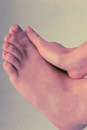
    The soft corn is caused by a section of bone in the fifth toe pressing against the bone in the base of the fourth toe. When this occurs, the skin between the toes is pinched excessively and becomes damaged. Because of moisture between the toes, the "corn" is soft. The skin is often white in appearance. On occasion, a small ulceration can form and the area can become infected. Surgical treatment for this condition is similar to the surgical correction for hammertoes.

    Description of the Surgery

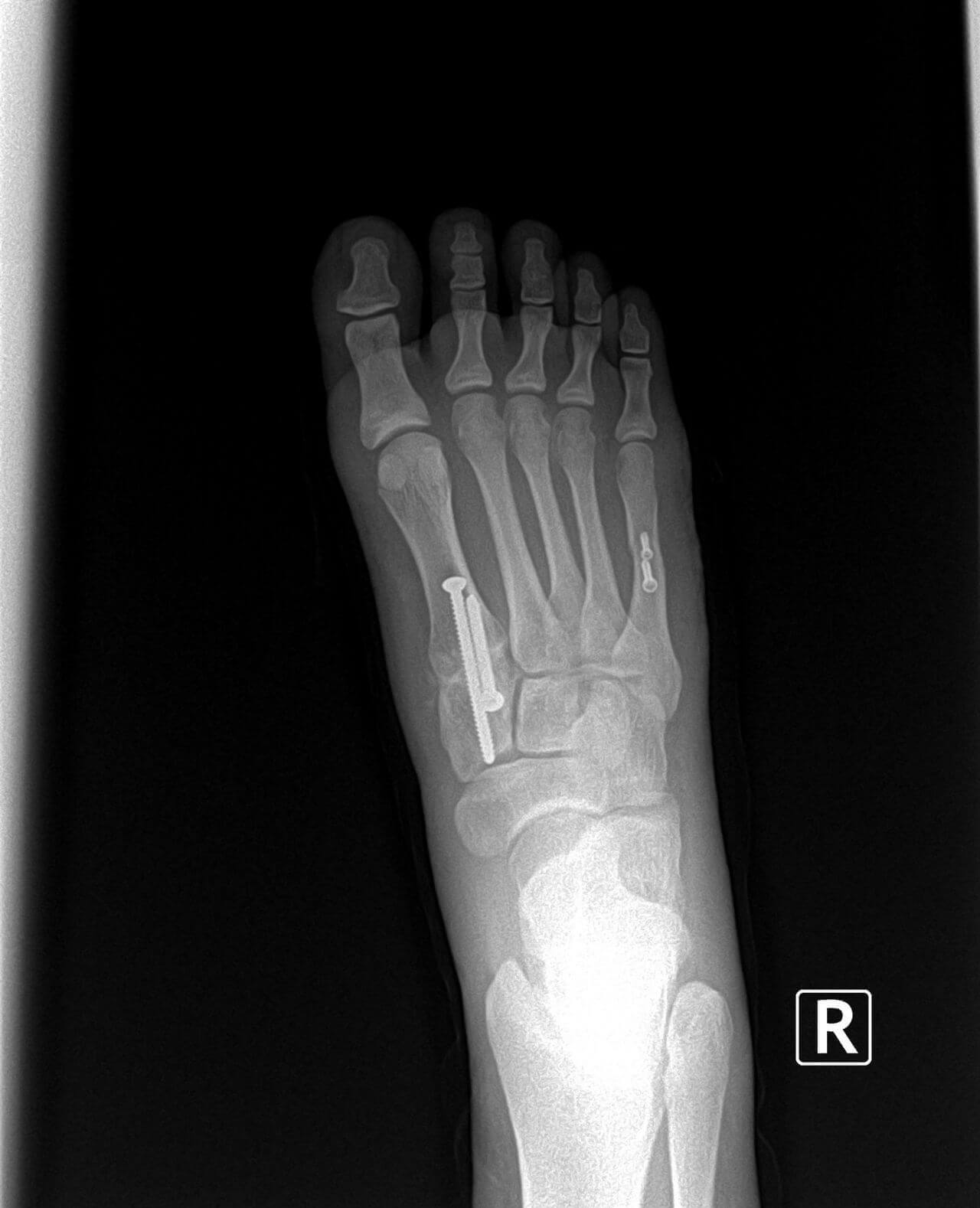
    An incision is placed on the top of the fifth toe. The ligaments about the joint in the toe are released to allow exposure of the head of the bone called the proximal phalanx. It is this section of the fifth toe bone that is pressing on the base of the fourth toe. The head of the proximal phalanx in the fifth toe is then cut and removed. If the toe is contracted or curled, then the tendon in the bottom of the fifth toe is released. On occasion, the surgeon will also make a small incision at the base of the fourth toe and smooth the bone in this area. In rare instances, the skin between the fourth and fifth toes may be so badly damaged that correcting the bone problem alone will not cure the soft corn. In this instance, the damaged skin between the toes must be removed. This procedure called a syndactylism necessitates removing the damaged skin between the toes and sewing the fourth and fifth toes together. This may sound like a drastic procedure, but it produces no loss of foot function. The procedure can be performed to leave a good cosmetic result. Surgery to correct soft corns can be performed in the doctor's office or in an outpatient surgery center. A local anesthetic is adequate for this surgery in most cases. Recovery from this procedure is much the same as for the correction of hammertoes. At the conclusion of surgery, a gauze dressing is applied.

    What to Expect Following the Surgery

    The patient should stay off their foot, keeping it elevated above their heart for a minimum of three days. Limited activity is recommended for two to three weeks. The stitches are removed in ten to fourteen days. During this period of time, the foot should be kept dry to reduce the risk of infection. A postoperative shoe is used to accommodate the bandage, and should be worn whenever the patient walks on their foot. Neglecting to wear the shoe while walking will cause the bandage to come loose, resulting in excessive movement at the surgical site, which will cause swelling, delays in healing, and possible infection. Once the stitches are removed, bathing the foot is permitted. Barefoot walking is discouraged for three weeks from the time of surgery. The patient should wear the postoperative shoe until they are able to wear normal shoes comfortably. This may take three to four weeks from the time of surgery. Some patients are able to return to normal shoes as early as two weeks.

    Possible Complications

    There are relatively few complications associated with this surgery. Excessive swelling which delays the healing process is the most common problem associated with the surgery. Postoperative infection rates are low but the risk increases if the surgical site is not kept dry. A reoccurrence of the condition is possible if enough bone is not removed or if the patient returns to wearing shoes that are too tight. If too much bone is removed then the toe may be floppy. It is normal for the toe to feel floppy for a few weeks following the surgery. The toe generally stiffens over time.

  • Surgical Correction Of Hallux Hammertoe
    The big toe, called the Hallux, is made up of two small bones called phalanges. This condition presents as a cocking up of the big toe at the joint between these two small bones. In the early stages of the condition the deformity is flexible, in later stages the deformity becomes rigid. It is caused by a variety conditions. Neurological diseases that cause muscle weakness or muscle imbalance in the muscles of the lower leg can result in the formation of Hallux hammertoe. This is commonly seen in patients after they have suffered a stroke or Cerebral Vascular Accident. Damage to certain areas of the brain during a stroke will frequently result in weakness and/or paralysis on one side of the body. If the stroke is not severe the patient may recover a majority of the function of the muscles in the legs and feet. However, a residual result may be a cocking up of the big toe.

    Other causes of the condition include damage or laceration to the tendon on the bottom of the big toe. Surgery to correct bunion deformities, in rare cases, may result in an imbalance of the structures about the big toe joint and cause the condition. An additional cause of hallux hammertoe is the absence of two small bones, called sesamoid bones, which are normally present beneath the big toe joint. There is an uncommon condition where a person may be born without these bones. More commonly however, the absence of one or both of the sesamoid bones is due to their surgical removal. In the course of correcting a bunion deformity one of the sesamoid bones may be removed. In another situation, a fracture of one or both of the sesamoid bones may result in the necessity to remove them to cure the pain associated with the injury.

    A high arched foot may also result in the formation of, not only a hallux hammertoe, but also hammertoes of all of the toes.

    A consequence of having a hallux hammertoe is irritation on the top of the toe from shoe pressure or the development of a painful callus on the end of the big toe. People who have had a stroke or who have diabetes with peripheral neuropathy may not have pain associated with the callus on the end of the toe. These areas may ulcerate and become infected.

    Diagnosis

    The diagnosis of hallux hammertoe is made by clinical exam. An x-ray is useful in determining the degree of the deformity and the condition of the joint. The presence or absence of the sesamoid bones is also made using an x-ray. If a neurological condition has not been identified and there is absence of trauma or previous surgery in the area, then evaluation by a neurologist may be appropriate.

    Treatment

    The need for treatment is based upon the level of symptoms the patient may be experiencing. Splitting the toe in an attempt to straighten it is of little value and is certain to fail. If treatment is needed, surgical correction of the deformity has the greatest level of success.

    If the deformity is flexible a simple tendon release procedure can be performed. This consists of making a small incision on the side of the toe and cutting the tendon in the bottom of the toe. If an ulceration or open sore is present on the end of the toe cutting the tendon to relax the toe may be all that is necessary to allow the ulceration to heal. This procedure can easily be performed in the doctor's office under a local anesthesia. Following the surgery a dressing is applied to splint the toe in a straightened position. The sutures and the bandage are kept in place for 7 to 10 days. The patient should keep their activities to a minimum during this period of time and keep the area dry. A post-operative type of shoe is worn to accommodate the bandage. Generally a patient can return to normal shoes within two weeks and resume complete normal activities in three weeks.

    If the deformity is rigid then fusion of the joint will be necessary to correct the deformity. Under certain circumstances the foot surgeon may elect to fuse the toe when the deformity is flexible. Fusion of the toe requires removing bone at the level of the joint in the toe. The articular surfaces of the joint are removed and the two small bones are abutted up against one another and held in place by a small screw. This fuses the two pieces of bone together resulting in permeate straightening of the toe. This procedure is generally performed in an out-patient surgery center or hospital. The surgery can be performed under a local anesthesia but the patient and surgeon may perfer to use a twilight anesthesia for the patient's comfort. Following the surgery a fluff dressing is applied. The sutures will remain in place for 7 to 10 days. During this period of time the patient should significantly reduce their activities and keep their foot elevated. It takes 6 weeks or longer for the bones to fuse. During this period of time the patient should wear a stiffed-soled post-operative type of shoe. Bending of the toe will delay or inhibit the fusion of the bones. Quite often it takes three months before the patient can return to full-unrestricted activity.

    Possible Complications

    Possible complications include infection, excessive swelling, and delays in healing or failure of the bones to fuse. Overall the procedure has a very high success rate. On occasion, over time, the screw may begin the cause irritation on the tip of the toe and have to be removed.

  • Surgical Correction/Arthritis In The Big Toe Joint

    Hallux limitus is a reduction in the ability to dorsiflex or move the big toe upward. This inability to move the big toe normally affects the way a person walks and runs. Eventually pain will develop in the joint behind the great toe. This joint is called the first metatarsophalangeal joint. This pain is due to an irritation of the joint capsule and/or destructive changes to the joint surfaces. As destructive changes to the joint surfaces become more severe, the toe gradually loses the ability to move upwards. This condition affects people of all ages and gender equally. There are numerous causes of hallux limitus. These causes include: arthritis both traumatic (ie: caused by injury) and systemic (ie: rheumatoid arthritis), an elongated first metatarsal (the long bone that is directly behind the big toe), an elevated first metatarsal, a first metatarsal bone that has too much motion, and a bunion deformity. All of the above conditions can have profound effects on the motion of the big toe joint. Identifying the cause will determine the various treatment options available to the patient.

    Diagnoses

    Diagnosis of hallux limitus is made by both physical and x-ray examination. Physical examination will reveal pain and limitation in the motion of the big toe. Pain is particularly severe with dorsiflexion or upward movement of the big toe. There is commonly mild swelling and bony prominences associated with the first metatarsophalangeal joint behind the big toe.

    X-ray examination of the foot will reveal the true severity of the patients condition. It will allow the physician to evaluate the joint for bone spurs, decrease in joint space, flattening of joint surfaces, and loose bodies in the joint. X-rays can also reveal the causes of hallux limitus such as an elongated or elevated first metatarsal.

    Surgical Treatment

    Surgical intervention is utilized when conservative therapy fails or the amount of deformity is too great. The goal of surgery is to obtain a more functional and less painful joint. Mild deformities are usually treated by removing bone spurs and prominences that develop around the first metatarsophalangeal joint. This helps to increase the amount of function and motion of the great toe. Occasionally, cuts in the bones called osteotomies, are made adjacent to the joint to correct for structural abnormalities. Osteotomies are held in place by screws, pins, or wires while the bone heals. By correcting for structural deformity the function of the great toe is increased as well as reducing the possibility for reoccurrence.

    Moderate to severe deformities require a more aggressive surgical approach. Moderate deformities are almost always treated with not only removing spurs that inhibit motion but also with osteotomies to realign the joint. Osteotomies are utilized to prevent progression to a more severe deformity. However, it is often difficult to determine in advance if adequate bony correction can be accomplished in order to prevent progression of the condition. Following the surgery the patient should wear a functional foot orthotic. These devices will correct much of the underlying functional cause of the deformity. When errosive changes in the joint result in absence of a large portion of the joint surface it may be necessary to perform a joint destructive procedure. These severe deformities require either a joint replacement or fusion procedure. The appropriate procedure depends on the patient's activity level and age.

    Recovery Time

    Your surgeon will usually require you to be off work for a minimum of one week. This is necessary to help control pain and post-operative swelling. Your return to work is dependent on the type of surgery that was performed and the demands of the job. Most patients are able to walk in a post-operative shoe or cast boot. Some surgeries however require the use of crutches and avoidance of placing pressure on the operative foot. At the end of one week most patients can return to work if they have a sedentary job. For patients that have an active job requiring a large amount of standing and walking, a longer recovery is necessary. Return to normal shoes and activities are dependent on the type of procedure and should be discussed with your surgeon. Following recovery from the surgery most surgeons will prescribe a functional foot orthotic. These devices are useful in reducing the reoccurrence of the condition and continued deterioration of the joint.

    Potential Complications

    Complications are rare, however they can occur in all surgeries. The most likely complications include: infection, delay or failure of the bone to heal, continued joint stiffness and pain, and prolonged swelling. Many complications can be avoided by taking prescriptions as directed and strictly following your surgeon’s post-operative instructions.

  • Surgery Of Hindfoot And Ankle Deformities

    Hindfoot and ankle deformity may result in decreased activity levels, inability to maintain meaningful employment, inability to walk or difficulty getting through activities of daily living. These severe deformities are often the result of previous trauma, congenital birth defects, acquired from degenerative changes throughout the course of one's adult life or may be secondary to systemic disease. Diseases such as diabetes mellitus, rheumatoid arthritis and various types of neuromuscular conditions may result in severe foot and ankle deformity. The deformities ultimately result in pain and difficulty walking.

    Non-operative therapy including orthoticsinjection therapy, anti-inflammatory medications, bracing, etc. are the first lines of treatment to provide comfort and maintain activities of daily living. However, certain deformities may fail to respond to non-operative care. Surgery can eliminate the deformity, decrease pain and increase activity level. Surgery is the ultimate solution to restore a relatively normal functioning foot that will permit a person to get through activities of daily living or maintaining meaningful employment.

    Surgical Intervention

    Surgery often consists of arthrodesis (fusion) procedures that permit a realignment of the existing deformity. The ultimate goal is to reconstruct the bony architecture of the foot so that the foot may be placed into appropriate footgear or bracing so that a patient can walk without pain. The exact surgery depends on the specific nature of the deformity. Realignment arthrodesis sometimes requires a 1-2 night stay in the hospital but is usually performed as an out-patient. These procedures are preformed under general inhalation anesthesia. Various types of internal and external fixation devices are often required either temporarily or permanently to maintain the foot or ankle in the appropriate alignment during the healing process.

    Recovery Time

    The postoperative convalescence includes three months in a non-weight-bearing short leg cast. This is followed by protected weight-bearing in a rocker bottom brace for an additional 2-3 months. The patient is then ultimately placed in custom molded or extra depth shoe with an appropriate orthotic or a permanent brace.

    Possible Complications

    Complications include infection, nonunion (failure of bones to heal together), fixation problems and malalignment (failure of realignment to be maintained). Additionally, stress fractures of the tibia (long leg bone) have been reported. These complications are managed by early recognition and prompt intervention. These complex procedures often require revisonal surgery to address complications.

    Summary

    Realignment arthrodesis of the hindfoot and ankle can be a very gratifying procedure. The ultimate goal is to have a patient who can walk without pain. Although surgery does provide realignment, some type of support in the form of an orthotic or brace is often required. The postoperative convalescence is quite extensive and the patient and their families should have a thorough consultation by the surgeon. It may take 6-12 months for a patient to get back to pre-surgical activity levels. A surgeon who is thoroughly experienced in hindfoot and ankle surgery should perform these procedures. The surgeon performing these types of procedures should have specialized continuing medical education, special postgraduate training and extensive experience with hindfoot and ankle surgery.

  • Plantar Fasciitis And Surgery

    Unfortunately, a small percentage of people that develop plantar fasciitis will not have success with conservative therapies.  We have an established treatment protocol for heel pain, making us a premier location for the treatment of plantar fasciitis.  The standard of care is six months to a year of conservative therapy before surgical intervention should be considered.  We have multiple surgical modalities available for patients with recalcitrant plantar fasciitis.  These surgical procedures can be performed with mild sedation in our outpatient surgery center that can save you both time and money by not having to go to the hospital or ambulatory surgery centers.  The procedures include:

    • gastrocnemius recession
    • plantar fasciotomies

    A gastrocnemius recession involves lengthening of the muscle in order to reduce the contracture.  A large percentage of people with chronic plantar fascial pain have limited range of motion of the ankle.  This limited range of motion often results in compensation via flattening of the arch, which leads to strain on the plantar fascia.  The post operative course requires a walking boot for about three weeks followed by a transition into proper shoe gear.

    The most common procedure for recalcitrant plantar fasciitis is a plantar fasciotomy.  This procedure involves lengthening a section of the fascia.  The idea is to reduce mechanical workload in the fascia thereby providing resolution of symptoms.  This procedure takes about 15 minutes and usually requires a period of non-weightbearing for about three weeks and an additional two to three weeks of protected weightbearing.   

  • Neuroma Surgery

    A neuroma is an abnormality of a nerve that has been damaged either by trauma or as a result of an abnormality of foot function. The most common location of neuromas is in the ball of the foot. In this area the nerve can become pinched and inflamed by the abnormal movement of the bones in the ball of the foot.

    As the condition progresses the nerve may become permanently damaged and surgical removal of the nerve may be necessary.

    The Surgery

    The surgical removal of forefoot neuromas is a relatively simple procedure. The surgery can be performed using a local anesthesia in the doctor's office or with intravenous anesthesia (twilight anesthesia) in an outpatient surgery center.

    Following administration of anesthesia, a skin incision is made on the top of the foot in the location of the neuroma. This is most commonly in the area between the second and third toes or between the third and fourth toes. An alternative surgical approach is to place the skin incision on the bottom of the foot in the location of the neuroma. Most surgeons perfer to make the skin incision on the top of the foot for several reasons. If the skin incision is placed on the bottom of the foot the patient may be required to use crutches for up to three weeks. Additionally, it takes longer for the skin on the bottom of the foot to heal. In the event that a thicken or irregular scar forms during healing, it may cause pain while walking. When the incision is made on the top of the foot the neuroma is easily found between the long bones (metatarsals) behind the toes. After the nerve is identified it is cut and removed. Once the surgery is completed a gauze dressing is applied. This bandage stays in place until the surgeon sees the patient on their first post-operative visit. On the first post-operative visit the surgical site is inspected and a new dressing is applied. The sutures are removed in 10 to 14 days following the surgery. During this period of time the foot must remain dry to reduce the risk of infection. The patient should limit their activities and keep their foot elevated above the heart as much as possible. A post-operative shoe is worn which allows the patient to do limited walking. The patient should not walk with out the post-operative shoe. Once the sutures have been removed the patient may bath the foot and attempt to wear a roomy stiff-soled walking shoe. It generally takes three weeks from the time of surgery before the walking shoe can be worn comfortably.

    Recovery Time

    The time required to be off from work will depend upon the type of work being performed and the type of shoe that must be worn. If the patient can work with their foot propped up and elevated with limited walking they may be able to return to work within a week of surgery. It is generally recommended that the patient not return to work until they can wear a normal shoe comfortable. Patients who have jobs that require prolonged standing, walking, kneeling or climbing may be off from work for as long as four to six weeks.

    Possible Complications

    The surgery has an over all high success rate, however, as with any surgery complications can occur. Possible complications include infection, excessive swelling, and delays in healing or continued pain. Also when a nerve is cut there is a small possibility that the nerve may grow abnormally producing a stump neuroma. If the patient walks on the foot more that what is recommended excessive swelling can cause bleeding or scaring that may result in continued pain and delays in healing.

  • Metatarsal Surgery

    There are five metatarsal bones in each foot. These bones are the long bones behind each toe. The metatarsal bone behind the big toe is called the first metatarsal. The metatarsal bone behind the little toe is called the fifth metatarsal. The most common metatarsal surgery is preformed on the first metatarsal for the correction of bunion deformity. The second most common metatarsal surgery is on the fifth metatarsal for the correction of tailor's bunion deformity. This article will address metatarsal surgery of the other metatarsals.

    Surgery on the remaining metatarsal bones is performed infrequently. When surgery is performed on the second, third, or fourth metatarsal bones, it is generally for the treatment of painful callouses on the bottom of the foot or for the treatment of non-healing ulcerations on the ball of the foot. Patients with rheumatoid arthritis may require surgery of the metatarsals, which is discussed in another section. Also surgery of the metatarsals may be necessary in instances of trauma of the foot where the metatarsal bones may have been fractured. This article will discuss elective metatarsal surgery.

    Painful callouses on the ball of the foot are due to an abnormal alignment of the metatarsal bones. If a metatarsal bone is lower than the others, excessive weight is placed on this area of the foot, and a painful callous may form. In people with diabetes, these areas of excessive pressure may break down and form open sores or ulcerations. Initial treatment generally consist of using a functional or accommodative orthotic to reduce the pressure to these areas. If this is not successful, metatarsal surgery may be considered.

    The Surgery

    The surgery consists of cutting the metatarsal bone just behind the toe. Generally, the bone is cut all the way through, and then manually elevated and held in its corrected position with a metal pin or screw. Following the surgery, the patient may be placed in a cast, or may be required to use crutches for several weeks. If a pin is used to hold the bone in place, it is generally removed in three to four weeks. Removal of the pin can be done in the doctor's office without the need for anesthesia. While the pin is in place, the patient should keep the foot dry to prevent infection. Generally it takes a total of six to eight weeks, or longer, for the bone to heal. During this healing period, the foot should be protected from excessive weight bearing. Walking prematurely on the foot can cause the bone to shift and heal in an incorrect position. This is the most common cause of failure with this surgery. Some studies indicate a failure rate as great as 60%. If the bone shifts downward, or is not elevated enough at the time of surgery, the painful callous may return. If the bone is elevated too much, a painful callous may form under the metatarsal next to the one which was operated on.

    Some surgeons will also cut out the painful callous on the bottom of the foot when they perform the metatarsal surgery. Rarely will a foot surgeon remove the painful callous without also performing the metatarsal surgery. Without correcting the metatarsal alignment, the painful callous is almost certain to return.

    Most surgeons prefer to do the surgery in an outpatient surgery center or hospital. In this setting, intra-venous sedation or general anesthesia can be used for the patient’s comfort.

    At the conclusion of the surgery, the surgeon places a gauze bandage on the foot. Generally, the bandage stays in place until the patient’s first follow up visit with the doctor. The skin stitches are removed in ten to fourteen days. If there are stitches in the bottom of the foot, they may remain in place for three weeks. The foot should be kept dry while the stitches and/or pin are in place to help prevent infection. May surgeons will have the patients wear a below the knee cast and/or use crutches for six to eight weeks. Other surgeons will allow the patient to wear a stiff-sole post-operative shoe, and allow limited walking on the foot.

    Recovery Time

    The time required to be off work will vary with the demands of the person’s job. A minimum of one week off work would be advisable with the patient staying at home with the foot elevated above the heart.

    Possible Complications

    Complications associated with this surgery are: infection, failure of the bone to heal in its correct position resulting in the return of the painful callous or transfer of the callous to a new location, delays or failure of bone healing, stress fractures of adjacent metatarsals, or excessive swelling. A common occurrence following the surgery is elevation of the toe associated with the elevated metatarsal bone that was operated on.

    Metatarsal Surgery to Treat Diabetic Ulcerations

    Diabetic patients with non-healing ulcerations on the ball of the foot may undergo a different type of metatarsal surgery. In this instance, the section of the metatarsal bone associated with the excessive pressure, called the metatarsal head, may be removed entirely. This is a relatively common and successful surgery that aids in the healing of the ulceration on the bottom of the foot. Possible complications with this surgery include infection, failure of the procedure to heal the ulceration, or the development of new ulcerations in adjacent areas on the ball of the foot. If the patient has poor circulation, further complications may be failure of the surgical sight to heal, or gangrene with partial loss of the foot or leg.

    In most instances of metatarsal surgery, the patient should use an orthoticin their shoe after the surgery has healed. This is especially important for the diabetic patient. The orthotic will reduce the risk of reoccurrence or the development of new areas of callous formation or tissue break down.

  • Heel Spur Surgery/Endoscopic Heel Spur Surgery

    Heel pain is caused by the excessive pull of a ligament in the arch of the foot called the plantar fascia. Generally the pain can be treated successfully without the need for surgery. However there are instances where non-surgical treatment of heel pain may fail and surgery may be necessary. Very often, the patient with heel pain will demonstrate a heel spur on an x-ray. In the past, many of the surgeries that were performed to relieve heel pain were designed to remove the bone spur. An incision was made on the side of the heel or the bottom of the heel, the spur identified and removed. Unfortunately, the healing time was very protracted and continued pain following the surgery was not uncommon. Now that the cause of heel pain is better-understood (See heel pain) the surgery is much more successful and the recovery time greatly diminished.

    The Surgery

    Most surgical procedures are aimed at detaching the plantar fascial ligament from its attachment into the heel bone. This may be accomplished with a small incision on the bottom of the heel or on the side of the heel. The procedure is performed by "feel". The surgeon inserted the scalpel blade and felt for the plantar fascia. Once they were confident that they had identified the plantar fascia they would cut the ligament free from the heel bone.

    In the mid 1990's a new procedure was developed called the "Endoscopic Plantar Fascial Release". This procedure developed by Dr. Steven Barrett DPM and Dr. Steven Day DPM of Houston, Texas uses technology similar to that used in arthroscopic surgery. A small incision is place on the side of the heel where a small cannula is placed allowing the insertion of an arthroscopic camera. The plantar fascial ligament is visualized and then cut using a small surgical blade. This allows the surgeon greater control during the surgery. The surgeon can control where and how much of the ligament is freed from the heel bone. Some surgeons still perfer to remove the spur at the time of surgery. This is the surgeon's choice and based upon their training and experience.

    The surgery is generally performed in an outpatient surgical center or hospital operating room. It can be performed under a local anesthesia, twilight anesthesia or a general anesthesia. At the completion of surgery a gauze dressing is applied and the patient placed in a post operative shoe or cast.

    Recovery Period

    Most patients are allowed to walk on the foot immediately but they are encouraged to limit their activities. The first week it is recommended that they stay off their feet except to use the restroom or have something to eat. After the first 3 to 7 days the bandage is removed and Band-Aids placed over the incision site. At this point the patient may attempt to wear a good supportive walking shoe if allowed to by their surgeon. Some surgeons perfer to protect the foot with a cast or have the patient use crutches. In 10 to 14 days the sutures are removed and the patient is allowed to bath the foot. It generally takes a minimum of three weeks before the patient is able to walk normally, with minimal discomfort. The patient should return to wearing their orthotics as soon as they are comfortable to wear in their shoes. Because the surgery does not address the cause of the heel pain (See heel pain) it is a good idea to wear orthotics following the surgery to reduce reoccurrence of the heel pain or other foot problems that might arise from excessive pronation of the foot.

    The amount of time needed to be off from work depends upon the demands of the job and the type of shoes that must be worn. If a limited amount of walking is required for the job and the patient is able to return to work wearing a cast the patient may be able to return to work in one week. If the job requires a lot of time standing, walking climbing or kneeling the patient may be required to be off work for three weeks or longer. These are general guidelines and it is important that the patient follow their doctor's instructions and guidance. Each surgeon has their own set of criteria to guide the patient through their surgery based upon their experience.

    Possible Complications

    Overall this surgery has a very high success rate, but as with any surgical procedure there are possible complications. If the patient walks excessively on the foot during the healing period following the surgery, it may damage the weakened soft tissues in the area of the heel. This can lead to continued pain in the heel or in other areas of the foot, particularly on the top and outside of the foot. This is due to jamming of a joint in the area. Infection is another possible complication. The risk of infection will increase if the surgical site gets wet while the sutures are in place. In some instances a continuation of the pain may occur. This can be due to the ligament not being cut all the way through at the time of surgery. Another cause of continued pain might be the presence on a pinched or damaged nerve called a calcaneal neuroma. A calcaneal neuroma is relatively rare and not easily identified and may only be recognized with the failure of the initial surgery. Some instances of failure of the surgery cannot be identified. In these cases the initial cause of the heel pain maybe due to something other than abnormal foot mechanics. Unfortunately, there are no tests for identifying these other causes of heel pain. They are generally diagnosed as a matter of exclusion, rather than with direct diagnostic testing.

  • Bunion Surgery - Shaft Procedures
    Hallux valgus or bunion deformities have may different surgical techniques for their correction. One group of procedures that your surgeon may use is the shaft osteotomies. These osteotomies are different from the head osteotomies and also the procedures performed at the base of the metatarsal or at the metatarsocuneiform joint, because they are performed in the middle of the first metatarsal.

    The shaft osteotomies were designed to use internal fixation (screws) and to correct larger deformities. In most of these cases, your surgeon will use 2 screws to fixate the osteotomy. The osteotomy is longer than the head procedures and has more inherent stability because of more bone contact. Also these procedures can correct larger deformities then the head procedures and about the same deformities as the base procedures.

    There are two basic shaft osteotomy procedures that your doctor may talk to about: The Z bunionectomy or the offset V bunionectomy. These osteotomies are very similar and are used interchangeably, based on different patient characteristics, by most surgeons that perform these procedures. The decision to use these procedures over other procedures is typically surgeon preference. In most cases, these procedures are used for patient with mild to severe structural bunions without hypermobility. In old patients with poor bone stock, the surgeon may opt for other procedures.

    What is the post-operative course?

    Typically, the patient is allowed to bear weight immediately after surgery in the a surgical shoe. Some doctors may have you use crutches for one to two weeks or use a slipper cast. This is surgeon's preference. It is not unusual for the front part of your foot to look bruised after the surgery. So at the first dressing change, do not be surprised if your toes and the top of your foot are bruised. This will dissipate in 3-6 weeks. At two weeks after surgery, the sutures are typically removed and at three weeks most patients are advanced into a surgical shoe. After the first or second week, your surgeon may have you start range of motion of your big toe joint. It is important that you follow your doctor's instructions on all range of motion exercises to help return motion to the operative foot. As with all surgery on the foot and ankle, the limiting factor to advance into different shoe gear is swelling. This swelling can last up from 6 months to one year after surgery. Typically most patients returned to pre-operative dress shoes in 6 to 8 weeks after surgery.

    With any surgery, complications are possible. Every procedure has unique complications and your surgeon will discuss these with you before surgery. Make sure that you ask any questions that you have about the surgery with your surgeon.

  • Bunion Surgery - Overview

    The surgical correction of bunions is dependent upon the severity of the deformity, the patient's over-all health and activity level.

    There are several different approaches to the surgical correction of bunions. Most commonly, the surgery is performed in the area of the big toe joint. The bony prominence is removed and the bone is surgically fractured to allow realignment of the joint and straightening of the big toe joint. This surgery is designed so that the patient can walk on the foot almost immediately following the procedure; however, activity must be significantly curtailed for several weeks following the surgery. Typically, the patient is instructed to remain home from work for at least one week with the foot propped up and elevated above the heart throughout the day. If the patient's job requires much standing or walking, they may be required to stay home from work for as much as six weeks. Often the patient may return to work sooner if they are placed in a removable below-the-knee walking cast. There are no short cuts to the healing time. Healing time is based upon basic physiological principles that are common to all human beings. Certain vitamins and nutrients may help with the healing process. Laser surgery does not alter the healing time and provides no significant advantage to the performance of the surgery.

    Surgical Correction of Severe Bunion Deformity

    If the bunion is more severe in nature, surgery is performed further back on the bone in order to straighten the big toe. When surgery is performed in this area of the bone, there is greater instability of the bone after it is cut and moved into a corrected position.

    The overall success rate and satisfaction of patients who have had bunion surgery is quite high. The most common complaint of patients is the healing time. This is particularly true if the patient is not adequately prepared or informed as to what to expect. Most patients experience minimal pain following the procedure and this pain is easily controlled with pain medication prescribed by the surgeon.

    Possible Complications

    Potential complications associated with the surgery are infection, over or under-correction of the bunion, joint stiffness, delays in healing or non healing of the bone, or healing of the bone in the wrong position. Most of these complications can be avoided by following the surgeon's instructions. Walking on the foot without the protection of a post-operative shoe or cast, or against the surgeons advice can lead to a dislocation of the bone where it has been cut. This results in delays in healing, non-healing of the bone or healing of the bone in the wrong position. Allowing the bandage to get wet increases the risk of infection. The most critical time for an infection to occur is within the first three days following surgery. Infection can also occur following this period of time but is less common.

    Joint stiffness following bunion surgery is common, but generally improves with time. Postoperative physical therapy is useful to improve the movement of the joint but is not always necessary.

    Place of Service and Anesthesia Considerations

    Most often the bunion surgery is preformed in an outpatient surgery center or hospital. However, at our office we are fully equiped with our own on-site surgical facilty. We are able to perfrom many procedures at our office. Because of our surgery center we save patients money, time and the frustrations associated with outpatient procedures. 

    Anesthesia for the surgery can range from a straight local anesthesia, given by injection into the area of surgery, to a general anesthesia with the administration of an anesthetic gas. A very common form of anesthesia is a combination of a local anesthesia and medicine given intra-venous to make the patient drowsy. This is commonly called twilight anesthesia.

    Generally there is very little blood loss during surgery. Most often the surgeon will use some form of tourniquet to stop bleeding during surgery. Because the surgery can be performed in a relatively short period of time the use of a tourniquet is very safe. Technically, the tourniquet can be left in place for as long as 90 minutes safely in most cases. The potential for the need for a blood transfusion with bunion surgery is nearly non-existent.

    Can My Bunion Come Back?

    It is important to understand that bunion surgery does not correct the cause of the bunion. Therefore there is the possibility that the bunion can reoccur. How quickly a reoccurrence will occur is difficult to predict. It may take several years or just a matter of months for the bunion to begin to come back. Bunions are caused by abnormal movement of a set of joints below the ankle joint in the foot called the subtalar joints. To help prevent the bunion from reoccurring the patient should be prescribed a functional orthotic. These are custom-made shoe inserts that correct the abnormal function of the foot. Generally they will fit in normal shoes without requiring the use of larger shoes.

    Bunions and tailor's bunions can be painful foot conditions that interfere with a patients ability to stay active and healthy. Bunions, also called metatarsus primus varus and tailor's bunions, also called a bunionettes can be treated conservatively or with surgery.  The causes of bunions are attributed to genetics, activity level, trauma and or shoe gear.

    A recent patient had visited the office with pain resulting from bunions. The paitent suffered from both a bunion and Tailor's bunion. Here are the preoperative x rays.


    Preoperateive x ray of the bunion and tailor's bunion.

    preoperative bunion and tailor's bunion


    The patient had a bunionectomy and tailor's bunion surgery corrected at our certified surgery center.  The patient was able to avoid the hassle and expense of a hospital, but with all the same comforts.  The patient required only intravenous sedation and was walking within one week after surgery.  The postoperative x rays can be appreciated below. 


    • The dramatic reduction of forefoot width can be appreciated on the x ray. 

    Bunion and tailor's bunion surgery, preoperative and postoperative x rays


    This represents part 2 of the Bunion and Tailor's bunion surgery


5 out of 5 stars
Total Reviews : 232