vendor

vendor

vendor

Wednesday, 10 August 2011 14:44

Heel Pain, Plantar Fasciitis And Orange Juice

When plantar fasciitis is causing heel pain, grab some juice and take a break.

Now, apply the juice directly to your foot...

If relief is what you are after and would like to try to ease the pain at home, try frozen juice.

Juice concentrates come in a cylindrical container that will easily roll along the bottom of your foot. When your plantar fasciitis is causing pain,  take a frozen juice concentrate. Right out of the freezer it may be too cold, so you may want to place a towel over the container. This will create a barrier so that the frozen juice container does not add to any discomfort. 

Before placing your foot on top of a frozen juice container check to see that it is sealed and do not apply too much pressure. The safety of your juice container and the floor below are at stake here. If the container is not sealed or is punctured you will likely have a sticky mess.

From a seated position you will gently roll the frozen juice container along the arch of your foot to your heel and back again. A few minutes of this can provide some relief to the pain associated with plantar fasciitis.

Heel pain that does not subside should be evaluated by a doctor. If you find that you are limping in the morning, when you first get out of bed, plantar fasciitis is the likely cause. Read more about the causes, diagnosis and treatment options for plantar fasciitis.

After rolling your feet over the juice concentrate you can easily mix up a beverage to enjoy and relax. Resting the painful plantar fascia is important. When the plantar fascia becomes irritated, inflamed or torn, it requires down time to heal. If possible try spending a little more time with your feet up and less of any activities that may have caused the strain of your plantar fascia.

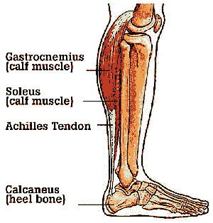
The plantar fascia is the thick ligament that connects your heel to your toes and is one of the most common causes of heel pain. Some of the common causes of this condition are a high arch, wearing high heeled shoes, running or being overweight. A tight Achilles tendon can also be to blame. Because of this stretching your calf muscles can relieve tension on your plantar fascia.

When the heel pain is frequent and nagging it is time to see a doctor. One common approach is to treat this type of heel pain is with prescription orthotics. Depending upon the severity of the injury (plantar fasciitis is generally caused by overuse, that causes strain and tearing of the plantar fascia) there are many treatment options. Surgery for plantar fasciitis is often the last resort.

Why use the rocker shoes?

The rocker style shoes tend to shift pressure off the heel and the forefoot. As a result, the rocking motion tends to decrease the stress load going to the midfoot.

What conditions can this help?

This can help heel pain problems such as plantar fasciitis and a thin plantar fat pad or cushioning on the bottom of the heel. This can also help forefoot problem such as pain at the forefoot joints-capsulitis, arthritis in the forefoot, Morton's neuroma, and a thin fat pad and the forefoot. This can also help arthritis and the midfoot with a rocker motion.

What problems could I experience from these shoes?

These types of shoes are inherently unstable, and for some people this is a problem. It often takes three weeks or longer to become accustomed to these shoes and you may feel a bit like you are "lurching around". You would have to be extremely careful going up and down stairs at least to begin with. The shoes can also cause more pressure in the midfoot, sometimes this causes some uncomfortable pressure in the arch but rarely the heel or forefoot. The shoes are not for everybody and it's important to get them from a store where you can use them for a “trial” period at home on the carpet, then they can still be returned if you don't get used to them or they are not comfortable.

Will they work with orthotics?

Yes, these rocker style shoes have removable insoles and work very well with orthotics. In general the combination of orthotics and rocker shoes gives the best possible combination of mechanical offloading to the symptomatic areas.

What about sandals?

The sandals are great, but they really accommodate orthotics. However because they are beneficial it is recommended that for some problems, you also get a pair of these rocker style sandals such as a sketcher or MBT sandals for summertime use.


Where to purchase the MBT shoes and sandals:

Where to purchase the sketcher shoes and sandals:


What is it?

neuroma is a benign thickening of the connective tissue or sheath around the nerve.  It commonly occurs in the 3rd interspace, betweent the 3rd and 4th toes.  However, it can occur in other locations.  The thickening of the nerve leads to irritaion which may cause perminant damage.

CausesMorton's foot type

The reason for development is unclear, but literature has related it to a person with a Morton's foot type.  That is an individual with a short first metatarsal which then leads to overload of the lesser metatarsals.  In addition, mechanical irritation and other forefoot pathologies like bunions and hammertoes have been atributed to the development of a neuroma.

Symptoms

Most patients relate a burning type sensation or electrical sensation radiating out the toes.  Others have described a numbness or sharp pain.  It is often worse in shoes and partially relieved with removal of the shoes and rubbing of the foot.  Still others relate the feeling as if they sock is balled up or something on the inside of their shoe.  The symptoms are usually progressive in nature.

Diagnosis

Physical examination of the foot in addition to imaging can provide the correct diagnosis.  Our office is equipped with real time diagnostic ultrasound which allows direct visualization of the neuroma. 

Non-surgical treatment

Treatment is usually initiated with conservative measures these include;

When is surgery needed?neuroma resection

Not very often for our patients we have a very high success rate with conservative care.  If surgery is needed it can be preformed in our ambulatory surgery center and takes only about 45 minutes.

Case study, example of a recent patient.

Recently we had a patient come to Issaquah Foot & Ankle Specialist with a 5 year history of neuroma pain.  The patient had tried multiple conservative modalities including; physical therapy, NSAIDS, topical medications, orthotics and cortisone injections.  The patient had been told the only recourse they had was surgical excision.  I discussed with the patient our technique for using alcohol injections and we proceeded with the series of injections.  They had a total of 5 injections and is now pain free and has returned to one of their favorite past times, walking .  

Alcohol Injections

I will briefly discuss the use of alcohol injections for neuroma pain.  The original article in foot and ankle journals describedthe use of 4% alcohol and required approximately 10 treatments.  The success rate reported in the literature was 89%.   We use approximately 25% alcohol and find we need to preform less treatments and have success that parallels the initial study.

Wednesday, 18 May 2011 14:33

Plantar Warts In Children

This is the last part of common condition we see in our pediatric patients

plantar wartWhat is it?

Warts are caused by the Human Papillomiavirus.  They can appear in any location on the foot but are common on the bottom or sole.  There are many different strains of the virus that produce warts but on the foot is is commonly associated with HPV strains 2, 4 and 6.  Warts can appear in clusters or as solitary lesions. 

Causes

Exposure to the virus itself causes one to contract the wart.  We see a correlation between patients who have sweaty feet or are exposed to moist environments, like avid swimmers or showers at the gym.

Symptomswart

Most patients feel a thickening of the skin or have pain with walking.  Sometimes one is able to view tiny black dots that are actually blood vessels to the wart. 

Diagnosis and Treatment

It is important to seek the diagnosis of a foot and ankle physician.  Warts are often confused for callus or other growths.  In addition treatments that work else where on the body often are not successful on the foot.  The goal of treatment is removal of the wart as quickly as possible.  We have great success with clearing of the warts in our pediatric populations. 

Heel pain in children and adolescence: Calcaneal apophysitis or Sever's disease is the most common osteochondrosis of the foot

Underlying anatomy: This is a condition that affects the cartilage growth plate and the separate island of growing bone on the back of the heel bone. This growth plate is called the physeal plate. The island of growing bone is called the apophysis. It has the insertion attachment of the achilles tendon, and it has the attachment of the plantar fascia.  This island of bone is under traction from both of these soft tissue tendon and tendon-like attachments. Severs disease resized 600

The causes of this problem: Mechanically, the heel takes a beating. And the apophyseal bone is located near the point of impact for the heel bone at heel strike and with most weight bearing activities. This includes running, jumping and walking. In addition to this, there is traction on this apophyseal bone and the associated physeal line of growth cartilage. This traction on the apopysis (island of bone) along with the impact of weight bearing activities can lead to inflammation and pain. Tight Achilles and calf muscles also can contribute to this problem.

Heavy impact activities like soccer, football and gymnastics are commonly associated with this problem.  I have seen this as early as with 8 year old boys and girls and as late as 14 year old teenagers. 

Additional factors: Having flatfeet or very pronated feet can make one prone to this mechanically. But also patient's have a very high arch foot structure tend to have a very high shock and high impact heel strike. This also puts extra stress on the heel and apophysis.

Symptoms typically include heel pain related to sports activities and worse after those sport and exercise activities.  But, some children who are not in a sport may also get this if they are physically active. The pain is usually on the back of the heel, the sides of the heel, the bottom of the heel, or a combination of all of these. We typically don't see swelling with this. Sometimes the pain is so bad the child will have to limp, or take a break from sports activity either for a few days or few months.

Diagnostic evaluation: This can include physical examination and x-ray evaluation. It is rare to require a CT scan for this. X-rays may show some increased density or sclerosis of the apophysis (island of bone on the back of the heel). This problem may be on one side or bilateral.

severs disease

Treatment: Involves a combination of rest, modified activity, anti-inflammatory treatments, shock absorption and support. In many ways, this is treated quite similar to adult plantar fasciitis. And in some cases there can be some inflammation of the plantar fascia. But this is not a typical finding.

Rest: If the problem is bad enough, it is important to totally rest the symptomatic foot.

  • Take a break from sport activity until the pain has significant improvement.
  • Severe cases will need to be treated with a cast boot.

Anti-inflammatory treatments:

  • Icing
  • Over-the-counter anti-inflammatories or as recommended by your pediatrician or podiatrist.
  •  Shock absorption and support
  • Don't go barefoot at home wear some type of good sandal or shoe
  • A significant and/or chronic case should be treated with prescription orthotics. This addresses mechanical problems that cause this problem.
  • Using an over-the-counter heel cushion inside of the shoe
  • Athletic foot taping

Stretching

  • Runners stretch to stretch out the calf muscle
  • A night splint will also help this.

Severe or chronic cases

  • Respond best to prescription orthotics with specific modifications for this problem.
  • May require a night splint
  • There are special daytime braces that may also help this.


At our clinic we are usually able to get our patients pain free within several weeks. Read our other post by Dr. Brandon Nelson about Sever’s Disease.

What is it?

ingrown nial

Ingrown toenails occur when bacteria gets trapped between your toenail and skin, usually near the borders of the toenail.  This causes a localized infection to occur which results in pain, swelling, redness and increased warmth.  There does appear to be a hereditary component to the development of ingrown toenails but, trauma, improper trimming and poor fitting shoes can contribute to the development as well. 

Treatment

We strongly discourage any treatment at home this can lead to increased infection and ultimately require more treatments to be preformed.   We have the newest techniques to minimize pain and discomfort while your ingrown toenail is treated.  We have treated thousands of ingrown nails and most of our patients have no recovery time after treatment.  We specialized in providing advanced foot and ankle care and recommend you seek a foot and ankle physician to treat this problem.  In addition, we will help provide tools and education to prevent the recurrence of another ingrown nail.


The picture below represents a painful ingrown nail before treatment.   

   before picture ingrown nail


The picture below represents a ingrown nail having received proper treatment at Issaquah Foot & Ankle Specialists.

after treatment


This diagram represents how a tiny piece of nail is removed to resolve the infection and pain.  The tiny piece of nail takes less than a minute to remove and most people experience very little discomfort.


What are you stretching?

Review from Part One

The system of the calf muscle and Achilles tendon heel bone or calcaneus, then plantar fascia is all connected. But this entire network the plantar fascia really doesn't stretch much, the heel bone and Achilles do not stretch and that leaves the calf muscle including gastrocnemius and soleus muscles. These are the primary target of our stretching exercises.

Various techniques for stretching

Weight bearing stretches:

Wall stretch: The typical runner stretch involves leaning up against a counter, a post, a wall or a tree and holding this position with the knee kept in a straight position (extended) and leaning forward until there is tension found in the calf muscle. Ideally the tension should be in the calf muscle and not the heel or the arch. Typically, this position is held for 15-30 seconds. If possible try to relax the calf muscle because it's more stretchable while it is relaxed. Then, bend the knee (slightly flexed) and repeat the stretch. You can alternate between the knee straight and slightly bent (extended and the knee flexed) and spent 3-5 minutes for the stretch. 

Pro stretch device. These are available online and in sporting good stores. These are a good tool to help you stretch more effectively.

Heel tilt stretch: You can do this standing beside a wall or a cabinet. Raise your forefoot up and positioned at an angle against the wall with your heel down on the floor and you lean towards the wall. Hold the stretch for 10-20 seconds and perform several of these.

Non-weight bearing stretches can include stretching first thing in the morning before getting out of bed. You can use a towel, jump rope or something to hang over the forefoot area and pull back and stretch gently holding this position for 10-30 seconds, repeating several times. This should not be painful but gently stretch out the calf.

Towel stretch: Before getting out of bed in the morning, take a towel and hook it over your forefoot then gently pulled back causing tightness in your calf. Hold the stretch for 10-20 seconds. Try to relax your calf as you do this. It should be firm but not cause pain.


How many stretches:

Spending several minutes over 4 times a day is optimal. It's hard to stretch too much- if you're warmed up prior to stretching.

Timing for stretching

Ideally you should stretch when you're body is warmed up. This could be after hot shower or hot bath or any time in the afternoon or evening. First in the morning your muscles are a bit tighter and may be less stretchable so you have to be more gentle.


There are many easy stretches that can help significantly with plantar fasciitis. These stretches do not require much time or special equipment. If you suffer from plantar fasciitis you can read more about this condition and other heel pain issues at our heel pain center.

Stretching is a great way to reduce the strain and pain caused by plantar fasciitis. However, it is important to know what to stretch and how to properly perform these exercises.

What are you stretching?

The system of the calf muscle and Achilles tendon heel bone or calcaneus, then plantar fascia is all connected. But this entire network the plantar fascia really doesn't stretch much, the heel bone and Achilles do not stretch and that leaves the calf muscle including gastrocnemius and soleus muscles. These are the primary target of our stretching exercises.

Anatomy – Stretching for plantar fasciitisThe plantar fascia spans from the heel to the forefoot and extends into the base of the toes. The calf muscle, including the gastrocnemius and soleus muscles, combine and attach at the heel to form the Achilles tendon. The soleus muscle attaches to the tibia and fibula below the knee and the gastrocnemius muscle extends and attaches above-the-knee.

It is important to think of the leg and foot including the gastrocnemius and soleus muscle as one functional unit. Or think of these muscles as one cable extending from above-the-knee along the back of the calf to the heel bone and then extending out to the forefoot. Of this entire complex, the most stretchable portion includes the gastrocnemius and soleus muscles.

The calf muscles are the most flexible components of this functional unit. As a result, this portion of this functional unit is where stretching is most important.


Why stretch?Leg muscles, stretching for plantar fasciitis

Walking, running and even standing in one place creates tension from the calf muscles that are attached and inserted into the heel bone. The plantar fascia extends from the heel bone to the forefoot and effectively completes this triangle. Therefore, there is always tension on the plantar fascia with any weight bearing activity and effectively it is the ligament of the bottom of the arch. When you're walking, the minute the heel comes up off the ground the plantar fascia becomes engaged and further tightens up as the great toe flexes upward during propulsion. As this action occurs in the plantar fascia, it tightens up and tends to help stabilize the arch. In some cases the arch height actually goes up. Therefore, the plantar fasciitis is clearly dynamic throughout the gait cycle and its functions very. But during many of these functions it has significant tension and traction at its origin site, the calcaneus. Individuals with tight calf muscles have more tension pulling on the heel bone and this effectively translates to tension and the fascia also.
 
For example, think of a woman who has worn high heel shoes her whole life. After period of several years, without stretching the calf muscle will tighten up and adapt to this high heel position. Therefore this individual has very tight calf muscles. So, when this particular individual goes from high-heeled shoes that they were most the time to a flat shoe or athletic shoe the calf muscle becomes quite a bit tighter. This additional tension will put more traction on the heel bone and effectively to the next connection which is the plantar fascia. Stretching may help compensate for this along with a night splint.

See more about plantar fasciitis and treatments.


Our next post (Part Two: Stretching for plantar fasciitis) will feature the different kinds of stretching techniques that can be used for plantar fasciitis.

Our long awaited summer is almost here. Below are five tips for keeping your feet, and your children's feet, healthy all season long.

Ankle sprains can result from improper footwear and we treat many ankle injuries in the summer. We also treat many cases of plantar fasciitis, and fit custom orthotics in the summer. With increased activity in the summer it is important to remain pain and injury free to fully enjoy our Pacific Nortwest summer. During a long winter like we experienced our feet and ankles can lose their natural strength. Our ankles and arches become accustomed to being supported in the sturdy footwear we wear in the winter months. As a result, our feet will need to be reconditioned to not having all of the support they used to.

Not all injuries can be prevented. However, consider these tips from the Issaquah Foot and Ankle Specialists to help keep you on your feet this summer.

Five of our tips for keeping your feet, and your children's feet, healthy this summer:

  • Transition slowly to less supportive footwear. Allow time for your feet to restore their strength to help avoid injuries this summer.
  • Apply sunscreen to your feet. Feet are at risk of sunburn also.
  • Protect your feet poolside. Avoid cuts, abrasions, bacteria, and viruses by wearing sandals or flip flops. This will help prevent athletes foot and plantar warts.
  • See a doctor quickly if you experience a puncture wound. If you step where you should not have and get a puncture wound have it treated properly. These types of injuries can lead to infection and other complications.
  • Wear the right type of footwear. Don’t wear running shoes for sports that involve a lot of side-to-side movement, such as tennis and basketball. 

Do not let foot and ankle injuries slow you down this summer. Custom orthotics can help keep you on your feet. Visit our heel pain center for more information about home remedies and treatment for the common causes of heel pain, including plantar fasciitis.

Bunions are a common presenting pathology for both adults and children.  Most patients notice an increase in the deviation of their great toe and often have difficulty in finding show gear.  A bunion is often described as a bump next to the big toe.  However, it is actually a structural malaligment of the the metatarsal bone. 

What you should know about bunion

Bunions are an inherited foot structure, they are a direct result of mechanical instability in your foot.  They are a progressive disorder, that is they will only get worse.  No tapping, splinting or stretching has been scientifically validated to reduce the bunion or reverse it.  Do not be fooled by websites selling splints or other devices to fix bunions, these are falsities.  Issaquah Foot & Ankle Specialists have specific treatment protocols establish from years of treating thousands of patients with bunions.  We have the ability to provide pain relief and minimize the progression of the bunion.  In addition we can correct the bunion in our surgery center if the need arises.  The correction is often preformed by cutting the bone and moving it into the correct position. 

preoperative bunion


On the above x-ray it is easy to appreciate the bunion if you look at the left side of the x-ray and look for the two small rounds bones at the end of the first metatarsal.  These two bones are called sesamoids and should be centered on either side of the metatarsal.  You can see how the first metatarsal has drifted so the sesamoids are shifted to the outside of the foot, or the right side of the x-ray.

post operative bunion

The above x-ray now demonstrates the correct alignment of the first metatarsal in relation to the sesamoids.  I preformed the above bunionectomy procedure in our ambulatory surgery center, it only took about 1 hour and the patient was walking the following day. 

5 out of 5 stars
Total Reviews : 236