vendor

vendor

vendor

Tuesday, 22 February 2011 11:19

Avoid Chilblains This Season

Many people are familiar with frostbite, but few have heard of chilblains, which are the mildest form of cold injury and the most common.  Chilblains occurs when the skin has been exposed to cold, wet and windy conditions and are seen more commonly in cold climates with high humidity. 

Symptoms of chilblains include:

  • Blistering
  • Small, itchy red areas on your feet
  • Swelling of your skin
  • Burning sensation on your skin
  • Changes in skin color from red to dark blue, accompanied by pain
  • Possible ulceration

Symptoms dissipate when the seasons change and warmer weather arrives, though they may recur seasonally for years.

There are many risk factors besides cold exposure that can increase your chances of getting chilblains.  They are as follows:

  • Being female
  • Being underweight
  • Location.  Chilblains actually are less likely to occur in colder and drier areas because living conditions and clothing used in these areas are more protective against cold.  People that live in an area with high humidity and low temperatures are are a greater risk for getting chilblains.
  • The time of year.  Chilblains are more common from November to April.
  • Improper fitting shoewear.  Chilblains can result from or be aggravated by unusual pressure on your skin, such as tight shoes.
  • Having poor circulation.  People with poor circulation tend to be more sensitive to changes in temperature, making them more susceptible to chilblains.
  • Having Raynaud's phenomenon. 

Chilblains can be treated with corticosteroid creams, blood pressure medication and infection prevention. 

We see many patients with chilblains this time of year, so please contact us if you are experiencing symptoms. 

Friday, 18 February 2011 11:16

Heel Pain Causes. What Causes Heel Pain?

The most common forms of heel pain are either mechanical or inflammatory or a combination of the two.

Note that we are talking about pain on the bottom of the heel and not the back. The back of the heel often includes Achilles tendonitis.

Mechanical types of heel pain include:

  • Plantar fasciitis
  • Heel spur pain
  • Thin fat pad, which results in a lack of cushioning
  • Tarsal tunnel syndrome or nerve entrapment at the heel
  • Calcaneal apophysitis in children (Severs disease)
  • Calcaneal stress fracture (heel bone fracture)
  • Sciatica or radiculopathy

Inflammatory or systemic causes of heel pain can include:

  • Gout
  • Rheumatoid arthritis
  • Reiter's syndrome and ankylosing spondylitis
  • Bursitis on the bottom of the heel between the plantar fat pad and the plantar fascia.

Typically, with either of these conditions there will be edema enlargement adjacent to the plantar fascia or involving the plantar fascia itself. Therefore, every step will be painful. Some of the inflammatory conditions such as gout can cause pain even when no pressure is placed on the foot. Classically, gout causes severe pain at nighttime when there is no weightbearing pressure.

Plantar fasciitis is one of the most common causes of heel pain. Typically, there will be edema and thickening of the fascia at its origin where it attaches at the heel bone. Without any weightbearing pressure and without the calf being utilized, the calf muscle tightens up. Therefore, pain is typically experienced first thing in the morning.  This is referred to as post-static pain or stiffness or post static dyskinesia. There are many treatments for plantar fasciitis to help address this pain in the morning.

A thin plantar fat pad (fat pad atrophy) is a common cause of heel pain. With a thin plantar fat pad, a patient is likely walking on the bone itself which means the periosteum, or outer layer of bone, really takes a "beating." After a full day of weightbearing pressure on this area without the anatomical plantar fat pad to cushion and protect the foot, the underlying structures become painful.

Heel bursitis causes heel pain because of its inflammatory component.  Bursitis is a fluid filled sac that puts pressure on the structures on the bottom of the heel. This pressure greatly increases with weightbearing pressure, therefore causing pain. Heel bursitis is an example of a condition that really has both inflammatory and mechanical components. This can start out as a "stone bruise" and progress to bursitis.

Tarsal tunnel syndrome is a form of nerve entrapment that can cause heel pain. A smaller medial calcaneal branch off of the posterior tibial nerve medial branch is usually more specific to heel pain.

Stress fractures can cause heel pain. There is often swelling with this condition. It should show up on an x-ray, but may require a CT scan or MRI for diagnosis.

Gout can also cause heel pain. It can occur at various locations in the body but it is very common in the foot, especially the great toe joint. The heel is in other locations and it can occur. Gout involves deposits of uric acid crystals. This is a breakdown product of protein that is normally process through the kidneys. It is usually is displayed as pronounced inflammation, redness and pain, especially at nighttime.

Rheumatoid arthritis can affect the heel, but more often affects the forefoot. This is an autoimmune disease.

Reiter's syndrome can cause heel pain, often at the achilles insertion but it can also target the plantar fascia attachment at the heel.  This condition usually involves the eyes and other mucous membranes.  Other similar conditions include ankylosing spondylitis and psoriatic arthritis.

Infections and neoplasms also can cause heel pain.

If you are experiencing heel pain, please don't hesitate to contact us

Thursday, 17 February 2011 11:13

Laser Bunion Surgery

Bunion deformities are related to a misalignment of bones in the foot. They are often an inherited foot disorder that can be exacerbated by activities, shoe gear and trauma.  Patients often  encounter advertisements for laser bunion surgery which guarantee less pain then traditional bunion surgeries. However, these are nothing more than a gimmick and no laser has the ability to correct a bunion deformity.

In order to correct the majority of bunions, bone must be cut and moved in order to realign the toe. This often can require a pin or screw to hold the bone in place once it is transposed.  Most bunion procedures can be performed in an outpatient surgery center, requiring no trip to the hospital.  Patients usually walk within the first couple of days and require minimal pain control.  Recovery from bunion surgery usually takes 6 weeks to 5 months depending on what procedure is performed. 

In addition to surgical correction of bunions, there are ways to slow down the progression of bunion deformities.  They are as follows:

After proper evaluation of a patient's specific situation, recommendations can be made. 

Tuesday, 15 February 2011 11:12

Prevent Injuries From Winter Activities

Nearly 50% of snow sports-related injuries are foot and ankle sprains.  Prevent injuries from winter activities before they start.  Follow these top five tips when you are out on the slopes to avoid injury:

  1. Choose your gear wisely.  The most critical piece of gear when skiing or snowboarding is a properly fitting boot, which helps you avoid blisters, sprains, shin splints and cold feet.  Pair those with high quality ski socks (not too thick) for maximum protection.  To ward off chilblains and frostbite, make sure to wear warm clothing and cover up your fingers, toes, ears and nose. 
  2. Stretch before and after your activity.  Many people overlook stretching before and after exercising, but it's imperative for winter activities due to the cold air.  Make sure you focus on your calves and toe flexors, as those will help protect your ankles and prevent injuries from developing.
  3. Know your ability.  If you haven't skiied before or haven't been physically active in a while, start slow.  Maybe take a few lessons before going out alone.  Even if you are a regular skiier, know your limits. 
  4. Keep hydrated.  Ironically, dehydration is more common in the winter months than in the summer.  Being well-hydrated helps ward off injuries while exercising.
  5. Always stay on marked trails.  Whether you are a seasoned veteran or new to the slopes, make sure to follow the marked trails only.  They are marked off for a reason and undoubtedly, the terrain beyond the ropes is very dangerous. 
Friday, 11 February 2011 10:25

Plantar Fasciitis Taping

Plantar fasciitis taping functions to reduce the stress and tension within the plantar fascia. The plantar fascia is considered the ligament of the arch and runs from the heel bone to the ball of the foot. Therefore, taping that follows the plantar fascia functions like an external splint. For the bottom of the foot, taping is very effective in that it allows the foot to still function in a fairly normal manner. This also greatly reduces the risk of tearing the plantar fascia. Many athletes tape their ankles or their feet to prevent injuries (or after an injury develops).

What you will need: 
You will need the tape to stick to the skin fairly well. If there is significant foot moisture or lotion on the skin, then this should be washed off. If the tape still does not stick well, you can wipe a thin layer of milk of magnesia on the foot.  Another option is to soak the foot in tea water. The tannic acid of the black tea is very good at toughening up the skin and also helps the tape stick more effectively.

  • A pair of scissors.
  • A roll of 1 inch tape (or 1.5 inch tape). We like to use Johnson & Johnson Zonas brand tape.
  • A roll of 2 inch tape (again 1.5 inch tape will work also).

What to expect from the taping: 
Taping allows individuals to do some of the normal athletic activities without "paying a price." However, it is best to use the tape as part of an overall treatment plan for the plantar fascia. Taping tends to offer temporary protection against further injury or progression of the plantar fasciitis and heel pain. Some people find more relief with taping than others. Some individuals find that initially the taping is very helpful but then the skin becomes irritated with the tape and they cannot tolerate ongoing taping.

The biggest concern: 
Skin irritation or allergic reaction. This can cause a severe blister or open wound for some individuals. Diabetic individuals or individuals with thin skin or peripheral vascular problems should not use tape. Once there is a slightest skin irritation, discontinue taping immediately and do not use the tape again. Instead switch to a PSC wrap, and other treatments.

Step-by-step instructions on LowDye taping method:

Step #1: The anchor strap: Wrap around the perimeter of the foot from behind the first metatarsal toward the heel, around the heel and then back toward the fifth metatarsal.

Step #2: Apply the first cross strap and tape across the bottom of the foot. Start just behind the first and fifth metatarsal-just behind the weightbearing ball of the foot. Apply the tape here and extend it up, overlapping the anchor strap on the side.

Step #3: Apply more of the cross straps. Overlap the next piece of tape by approximately 50% and continue taping layer by layer back toward the heel. It is not necessary to cover the bottom of the heel. Usually there are 3-4 cross straps.

Step #4: Finally, lay another piece of tape just over the anchor strap to cover all of the unfinished ends of the tape along the perimeter of the foot from the first metatarsal back around the heel extending to the fifth metatarsal.

Step #5: The last optional piece of tape would be to wrap a piece of tape all the way around the forefoot similar to the very first crosspiece in step #3.

Variations on the LowDye taping technique:
You can have a piece of 1 inch tape forming an "x" just in front of the bottom of the heel near the area of the pain. This can be done between steps #3 and #4 above.  Another option is moleskin placed in the mid arch. This is also called a "T. strap."

Medical disclaimer: This information is not intended to provide medical advice or treatment. It is important to realize that there are potential risks with applying tape. As mentioned, one would be an allergic reaction. Another would be skin irritation or blister formation. In some individuals, these blisters could develop serious infections. Therefore, it is best to check with a medical doctor before using tape on your skin.

Wednesday, 09 February 2011 10:19

Dry Needling For Plantar Fasciitis: Part 3

New! Modification of dry needling for plantar fasciitis procedure. We are not aware of any other clinic or facility that is providing this treatment other than our clinic at this time.

One of our goals with dry needling has been to use the smallest dose of cortisone possible. This is because there is a small risk of rupture of the plantar fascia with any cortisone injection. The way that this risk has been reduced dramatically has been to inject between the plantar fascia and the fat pad (and avoid the plantar fascia itself). It is possible to do this procedure without using any cortisone at all. Some doctors have the theory that the use of cortisone with dry needling is counterproductive. This is because cortisone inherently decreases inflammation and the dry needling procedure itself is designed to actually allow for some inflammation and release of platelets with associated growth factor.

With this in mind, we are starting to do this procedure for some individuals using platelet-rich plasmainstead of cortisone. The goal is to further augment the growth factor concentration after this procedure and allow the treated plantar fascia the maximum healing potential possible without adding cortisone which inherently decreases inflammation.

Platelet-rich Plasma Procedure

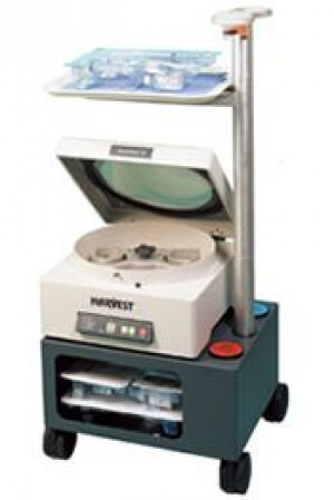
This procedure is very similar to typical dry needling protocol. A local anesthetic is used. We again use our electrical nerve stimulation to help minimize discomfort from the local anesthetic. Then there is no discomfort whatsoever. 20 cc of blood is drawn from the patient and put in a two-stage centrifuge. A high-speed centrifuge is able to separate out the component of blood that contains the platelets. So for the 20 cc of blood, we now have obtained 3 cc of platelets (with associated growth factor). This growth factor is then used instead of the cortisone. It is injected at the interface between the plantar fat pad in the plantar fascia. And we also inject some of this into the plantar fascia itself.

There are several different local biotech companies that help with the preparation of the platelet rich plasma. They typically charge the patient a direct fee for this service that is not covered by insurance. However, the doctor’s procedure for the ultrasound-guided injection is typically covered by insurance.

The post procedure protocol is essentially the same as typical dry needling. Some of the elements of this include the use of a cast boot and avoiding anti-inflammatory medication.

Thursday, 03 February 2011 03:10

Plantar Fasciitis And Night Splints

Plantar fasciitis can be treated in many different ways and you should always come in to see us so we can devise a treatment plan together before you try any home remedies.  

With that being said, one of the treatments for plantar fasciitis the use of night splints.  A night splint is a brace that attaches to not only your foot, but also the ankle and the lower leg.  It's worn at night when you go to bed and its intended purpose is to stretch the plantar fascia ligament while you sleep. 

The purpose of stretching out your plantar fascia is so the stress on the inflamed area of your foot is minimized.  Some studies have shown that night splints actually help reduce the pain that plantar fascia patients feel when they initially get out of bed in the morning.  They also have been known to reduce a patient's pain level throughout the day as they spend more time on their feet while working and performing everyday tasks. 

In addition to night splints, orthotics are another treatment option for patients with plantar fasciitis.  Both night splints and orthotics can treat many more disorders than just plantar fasciitis.  These include, but are not limited to:

With that being said, there are many additional treatment options for plantar fasciitis and in order to find out which treatment is best for you, please contact us to make an appointment. 

Wednesday, 02 February 2011 10:11

Dry Needling For Plantar Fasciitis: Part 2

What to expect
At Issaquah Foot and Ankle Specialists, we have developed a treatment protocol and have fine tuned the technique for dry needling for plantar fasciitis. In addition, we have developed adjunctive treatments that help further ensure that chronic plantar fasciitis is resolved. Dr. Young has personally communicated with Dr. Lucia Sconfienza by e-mail soon after the results were first presented. He discussed details of the procedure to make certain that we were able to provide our patients with the best possible treatment and outcome.


Who is a candidate for dry needling
Those individuals who have already tried other means of treatment for plantar fasciitis including but not limited to prescription orthotics, excellent shoes, a night splint, cortisone injections, taping and bracing.

Diagnostic ultrasound imaging typically will show enlargement of the plantar fascia in those individuals with severe plantar fasciitis. The typical thickness of the plantar fascia at the heel is anywhere from 3 to 4 mm thick maximum.  Those individuals with chronic plantar fasciitis typically will be anywhere from 20% to even 100% thicker than normal. This means that the fascia can be 5 mm to 8 mm thick (even up to 10 mm in some severe cases). If an individual does not have significant swelling (edema) of the plantar fascia, then the prognosis for improvement with dry needling and many of the other traditional plantar fasciitis treatments tend to be less effective. In general, it is a severe case without swelling of the fascia and there are other significant causes of the heel pain instead of plantar fasciitis. 


Dry needling description

First, we utilize our electrical stimulation in conjunction with the local anesthetic to help minimize discomfort.  After the local anesthetic has fully taken effect, the procedure is done with no discomfort whatsoever.

High-resolution ultrasound guidance is used to direct the procedure, which allows for the precise finding of the exact location. A needle and syringe is used to repeatedly probe the fascia throughout the thickest portion at the calcaneal attachment site.  This probing can go all the way down to the attachment and periosteum (outer layer bone). Once this has been done for the prescribed time frame, then we typically utilize a combination of Kenalog and dexamethasone phosphate at the interface between the plantar fascia in the plantar fat pad. This is another example of where the ultrasound guidance is essential. Using this technique, we can make certain that the cortisone is not injected into the plantar fascia itself or the plantar fat pad. Injecting into the plantar fascia would raise the risk of a partial tear or rupture of the plantar fascia. In addition, injecting into the plantar fat pad can cause long-term thinning (atrophy) of the fat pad.


New treatment variations on this technique
This is where instead of using cortisone, we use autogenous platelet rich plasma with growth factor. It is also possible to do this procedure without using any cortisone.


Frequently ask questions
Is it safe?
Yes, dry needling is very safe.  There are few risks associated with the procedure and these risks are minimal compared to surgical risks.  A patient may experience an allergic reaction from the anesthesia and they do run a small risk of post-injection infection.  Some patients may have pain for a few days following the procedure as well. 

Is there any pain with the procedure?
No, a patient shouldn't experience any pain during the procedure.  We use a local anesthetic to numb the foot as well as electrical stimulation with our TENS unit to make sure the procedure is pain-free. 

How long have you been doing dry needling? 
Our clinic started providing the service in December of 2008. Our clinic has been providing high-resolution ultrasound guided injections for over 5 years.

Does my insurance cover this procedure? 
We accept many different insurance plans at our office.  This is something that our billing staff can help you with.


Post-procedure treatment protocol essentials:

  • Avoid anti-inflammatory medication and icing
  • Use a cast boot.  This is done to protect the soft tissue in the heel as it now starts the healing process.
  • Allow for bruising and inflammation.  When you tear or sprain a tendon or ligaments, there is deep internal swelling or bruising that occurs due to the need to protect the tissue which allows for soft tissue healing phases to occur. The same should occur with this procedure.

What else can be done to help make sure this is effective:

  • Use a night splint every night for the first 2-3 months.
  • Prescription orthotics that are specific to your problem and to plantar fasciitis. You want to treat the mechanics that caused the problem to begin with.
  • Electrical stimulation may be beneficial to the post-procedure healing time frame.


Contact us today for an appointment or to learn more.

Cortisone injections are commonly used to help treat the short-term heel pain caused by plantar fasciitis. This course of treatment is decided upon after we have assessed the patient and their specific situation, so it's not a treatment we give to everyone suffering from plantar fasciitis.  The intention of the injection is to help minimize the pain by injecting their cortisone directly into the plantar fascia, under the heel or on the big side of the heel or arch. 

One of the biggest benefits of this procedure is that it is a non-surgical option to helping heel pain.  Surgery can be very invasive and rather costly when done at a hospital.  We do have a surgery center here in our office for our patients that need it, which saves both time and money.  As for the cortisone injections, some people see a noticeable decrease in their heel pain after their injection. 

Some patients might be leery of an injection in their foot, but our office has a solution to this fear.  We used what is called a Transcutaneous Electrical Nerve Stimulation (TENS) unit. The TENS unit works by sending stimulating pulses across the surface of the foot and along the nerve strands.  The stimulating pulses help prevent pain signals from reaching the brain so a patient doesn't even feel the injection as it's happening. 

If you are experiencing heel pain, please contact us to make an appointment. 

Wednesday, 26 January 2011 10:05

Plantar Fasciitis Home Remedies

When you are experiencing pain causing by plantar fasciitis, it is always best to come in to see us so we can assess the situation and provide the proper course of treatment.  However, there are a few home remedies that can help in relieving your pain in the meantime.

  • Cut back on your exercise routine.  Exercise is a very common trigger of plantar fasciitis, so if you are an athlete, try cutting back on your routine.  Decrease the number of miles you are running or the amount of time you are spending on the cardiovascular machines at the gym.  This can make a huge difference in the amount of pain you are feeling.  When in doubt, stop your exercise routine altogether until you come in to get assessed. 
  • Switch up your exercise routine.  In addition to cutting down your routine, opt for lower-impact exercises to alleviate the stress on your feet.  High-impact exercises such as running and aerobics put a lot of strain on your feet, which increases your heel pain, so avoid those types of exercises as much as possible.  Instead, try activities such as swimming or an elliptical machine, which are both low-impact exercises that won't cause your heel pain to flare up. 
  • Stretch your arches.  To help decrease the pain in your heel, get into the routine of performing simple stretches daily.  These can be done in the comfort of your own home and don't take but a few minutes of your time.  Stand and stretch your arch using the assistance of a household object or stretch your arch while sitting.  Either option will help alleviate your pain.
  •  Ice your feet.  Applying an ice pack to the bottom of your feet will help to decrease the inflammation caused by plantar fasciitis.  You can perform this home remedy for 15 to 20 minutes 3 to 4 times daily.  Another trick that works well is freezing water in a small paper cup.  Once it's frozen, you can peel the lip of the cup away and massage your foot with the ice.  This also is a great solution for shin splints.
  • Stay off of your feet. We know how hard it is on a day-to-day basis to stay off of your feet, but this will really help the pain and inflammation caused by plantar fasciitis.  Rest as much as possible and when you can, use a pillow to elevate your feet and relax. 
  • Massage with a golf ball.  This may sound strange, but many patients have found the golf ball massage to be the best home remedy for their plantar fasciitis pain.  Use the golf ball to massage the bottom of your foot, which stretches the plantar fascia and decreases adhesions in the area.   

As mentioned before, none of these home remedies should take the place of a visit to our office.  Every patients' case of plantar fasciitis is different and some may need more extensive treatment than those remedies offered above, but use these in the meantime to gain instant relief from your pain. Contact us today for an appointment.

5 out of 5 stars
Total Reviews : 240