Washington Foot & Ankle Surgery Center

  • Morton’s Neuroma – Surgery Alternatives

    Have you ever felt like your sock is bunched up under the ball of your foot, or like you have a stone in your shoe, along with a burning sensation in your toes? This is likely caused by a Morton’s neuroma.

    Many people believe that this pain will go away after time. However, after prolonged suffering many people believe they must live with this pain. With Morton’s neuroma these beliefs are not true. It is very unlikely that the pain from a Morton’s neuroma will pass without intervention. The condition can be treated by several methods; we have previously discussed Morton’s Neuroma Treatments.

    This condition is a thickening or enlargement of the nerve tissue in the ball of the foot. Neuromas often form when the nerve is ir­ritated or compressed. Sometimes runners and joggers develop them because of the repeated pres­sure that occurs when feet hit the pavement. Women can be espe­cially at risk, because running on hard, paved surfaces and then switching to high heels with nar­row toes places a lot of stress on the feet. Most neuromas begin gradually, and you may be able to eliminate the pain temporarily by massaging your foot, changing footwear or a rest from running. Unfortunately, neuromas tend to worsen over time as the tempo­rary changes in the nerve become permanent.

    Early intervention is important. Surgery is often performed to address this condition. However, there are alternatives to surgery for Morton’s neuroma. In fact a number of our patient's recently received dehydrated alcohol injections and are doing exceptionally well. Statistically the success rate has even been slightly higher with these types of injections opposed to surgery. We use high definition ultrasound imaging to guide the injection directly to the painful area. We can also incorporate nerve stimulation to significantly reduce any sensation associated with the injection. Research has indicated that success rates from surgical treatments are between 80-85%. The latest research of dehydrated alcohol injection therapy for Morton’s neuroma has been 89%. Therefore we believe we are well on our way to ending the pain associated with Morton’s neuroma with less invasive treatments. Interested in treating your Morton's neuroma with the virtually pain free dehydrated alcohol injections guided by ultrasound imaging we provide? Request an appointment today!

    Read more information about Morton's Neuroma Treatments.

  • Cartiva Implant

    Cartiva Implant

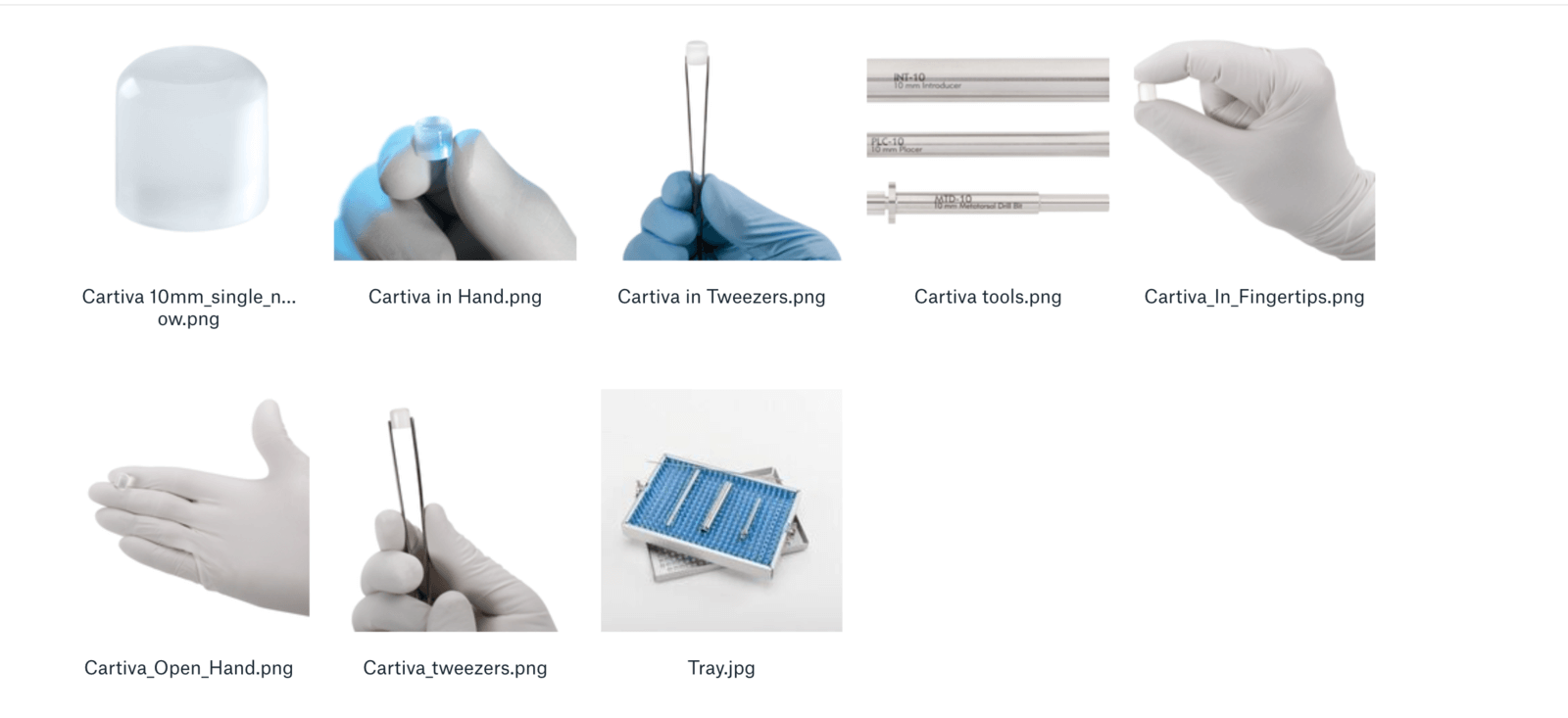
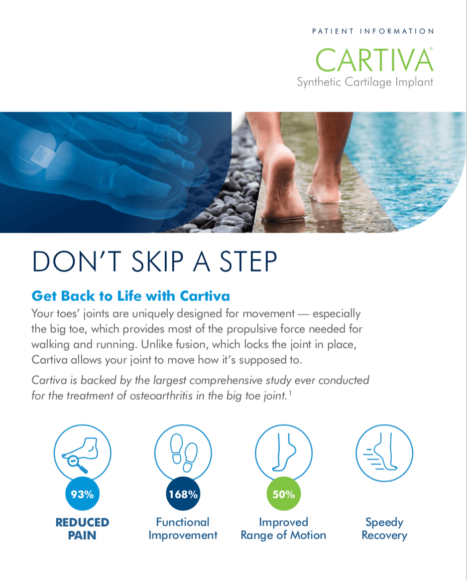
    A Cartiva Implant is used to treat arthritis in the joint of the big toe. When you have arthritis, the cartilage tissue in the big toe wears down. Cartiva is made of a polymer that is biocompatible, durable, and organic -- similar to how natural cartilage is. We specialize in Cartiva Implants here at Issaquah Foot & Ankle Specialists.

    For more information on Cartiva Implants, please read the Patient Information Brochure.

    In addition, make sure to check out the Frequently Asked Questions concerning Cartiva Implants.

    Cartiva Implants have been found to be safe and effective, as many peer reviewed studies have shown.

    Testimonials


    Cartiva In The Media


    For more videos on Cartiva, be sure to check out our Youtube channel.

  • Ankle Fractures
    A fall, an accident, or a hard blow may cause one or more bones in the ankle to fracture or break. Depending on the type of injury, an ankle may fracture in several ways. Spiral fractures may result from twisting injuries. If bone pulls away from bone, the break is often straight. After a bad fall, or a heavy blow, bone may be crushed. Although ankle fractures tend to cause pain, you may be able to walk on the ankle. However, this is not a good idea. Your physician should check any injured ankle.

    Treatment

    Your treatment depends on where and how badly your ankle has been broken. Your physician will feel your ankle and foot for swelling and any displaced bones. He or she may also check to see if the ankle joint still moves. Depending on how the injury occurred, your physician may examine your leg from the knee down. X-rays will be taken to show the type of break, its exact location, and the extent of the damage.

    After a fracture, a cast may be used to hold the bone in its proper position for healing. Sometimes the sections of broken bone must first be realigned. This is called reduction. The type of reduction is based on how far the bone has moved from its normal position.

    Closed Reduction

    If you have a clean break with little soft tissue damage, closed reduction may be used. Before the procedure, you may be given a light anesthetic to relax your muscles. Then your doctor manually readjusts the position of the broken bone.

    Open Reduction

    If you have an open fracture (bone sticking out through the skin), badly misaligned sections of bone, or severe tissue injury, an open reduction will be used. A general anesthetic may be used during the procedure to let you sleep and relax your muscles. Your doctor then makes one or more incisions to realign the bone and repair soft tissues. Screws or plates may be used to hold the bone in place during healing.

    Casting a Fracture

    To make sure the bone is aligned properly, an x-ray is taken. The ankle is then put in a cast to hold the bone in place during healing. You will probably have to wear the cast for 4 to 8 weeks. For less severe fractures, a walking boot, brace, or splint may be all that is needed to hold the bone in place during healing.

    Once your fracture has been treated, your physician will tell you how to help it heal. You may be told to limit your use, take medications, and elevate the foot. If you have a cast, remember to keep it dry. To reduce swelling and control pain, elevate the ankle above the level of your heart. This simple action can help control symptoms throughout your recovery.

    Protect your ankle by giving it enough time to heal. When you do become active again, expect some swelling and stiffness. To build strength and help avoid re-injury, your physician may prescribe physical therapy or home exercise. Ankle support may also help. Exercise increases the flexibility of your ankle. If your physician agrees, try the following exercise. Use your foot to write out the alphabet in the air. Be sure to work from the ankle and foot, not from the knee.

  • Surgical Treatment Of Adult Aquired Flatfoot
    Adult acquired flatfoot deformity or posterior tibial tendon dysfunction is a gradual but progressive loss of ones arch. The posterior tibial muscle is a deep muscle in the back of the calf. It has a long tendon that extends from above the ankle and attaches into several sites around the arch of the foot. The muscle acts like a stirrup on the inside of the foot to help support the arch. The posterior tibial muscle stabilizes the arch and creates a rigid platform for walking and running. If the posterior tibial tendon becomes damaged or tears the arch loses its stability and as a result, collapses causing a flatfoot. Adult flatfoot deformity can occur in people of all ages and gender however, it occurs most commonly in sedentary middle aged to elderly females. There are several risk factors for posterior tibial tendon dysfunction that include: obesity, steroid use, systemic inflammatory diseases such as rheumatoid arthritis, trauma, being born with a low arch, and diabetes. It occurs most commonly in one foot however, it can occur in both feet especially in people with systemic diseases such as diabetes and rheumatoid arthritis.

    There are four stages of posterior tibial tendon dysfunction. In the first stage the posterior tibial tendon is inflamed but has normal strength. There is little to no change in the arch of the foot. In stage two the tendon is partially torn or shows degenerative changes and as a result loses strength. There is considerable flattening of the arch without arthritic changes in the foot. Stage three results when the posterior tibial tendon is torn and not functioning. As a result the arch is completely collapsed with arthritic changes in the foot. Stage four is identical to stage three except that the ankle joint also becomes arthritic.

    Surgical Treatment

    Surgical correction is dependent on the severity of symptoms and the stage of deformity. The goals of surgery are to create a more functional and stable foot. There are multiple procedures available to the surgeon and it may take several to correct a flatfoot deformity.

    Stage one deformities usually respond to conservative or non-surgical therapy such as anti-inflammatory medication, casting, functional orthotics or a foot ankle orthosis called a Richie Brace. If these modalities are unsuccessful surgery is warranted. Usually surgical treatment begins with removal of inflammatory tissue and repair of the posterior tibial tendon. A tendon transfer is performed if the posterior tibial muscle is weak or the tendon is badly damaged. The most commonly used tendon is the flexor digitorum longus tendon. This tendon flexes or moves the lesser toes downward. The flexor digitorum longus tendon is utilized due to its close proximity to the posterior tibial tendon and because there are minimal side effects with its loss. The remainder of the tendon is sutured to the flexor hallucis longus tendon that flexes the big toe so that little function is loss.

    Stage two deformities are less responsive to conservative therapies that can be effective in mild deformities. Therefore, these patients may be candidates for a 15 minute outpatient procedure to correct the flexible flatfoot deformity which is referred to as hyperpronation. The procedure is called a Subtalar Arthroereisis. It involves the placement of an implant in the space under the ankle joint (sinus tarsi) to prevent only the abnormal motion, but still allowing normal motion. This brief procedure only requires very little recovery time, and is completely reversible, if necessary. Your surgeon can consult you about this exciting, life-changing procedure, or more information can be obtained at www.hyperpronation.com.

    Stage three deformities are better treated with surgical correction, in healthy patients. Patients that are unable to tolerate surgery or the prolonged healing period are better served with either arch supports known as orthotics or bracing such as the Richie Brace. Surgical correction at this stage usually requires fusion procedures such as a triple or double arthrodesis. This involves fusing the two or three major bones in the back of the foot together with screws or pins. The most common joints fused together are the subtalar joint, talonavicular joint, and the calcaneocuboid joint. By fusing the bones together the surgeon is able to correct structural deformity and alleviate arthritic pain. Tendon transfer procedures are usually not beneficial at this stage.

    Stage four deformities are treated similarly but with the addition of fusing the ankle joint.

    Recovery Time

    Time off work depends on the type of work, as well as the surgical procedures performed.

    Potential Complications

    Complications can occur as with all surgeries, but are minimized by strictly following your surgeon?s post-operative instructions. The main complications include infection, bone that is slow to heal or does not heal, progression or reoccurrence of deformity, a stiff foot, and the need for further surgery. Many of the above complications can be avoided by only putting weight on the operative foot when allowed by your surgeon.

  • Surgical Removal Of Giant Cell Tumors

    This tumor was once thought to be a cancer of a tendon sheath. It is now known to be a benign non-cancerous tumor of a tendon sheath. These masses are generally found on the toes, top of the foot or sides of the foot. They are always closely associated with a tendon sheath. They can also occur deep inside the foot. They slowly enlarge but never grow any larger than 4cm in size. They are firm irregular masses that are commonly painful. The pain seems to be a result of the tumor pressing firmly on the surrounding tissues and due to the interference with the function of the tendon the mass is growing from. As the tendon grows it can press so firmly on the bone it lays next to, that it can cause erosion of the bone. It is because of this erosion of bone that the tumor was once thought to be cancerous. Cancerous tumors can have the characteristic of invading bone through aggressive and destructive means. The erosion of the bone associated with giant cell tumors is due to pressure on the bone and not due to the invasion of the bone by the tumor. Other common soft tissues masses that may occur in the foot are ganglions, fibromas.

    Diagnosis

    The diagnosis of a giant cell tumor is generally made by a pathologist following removal of the mass. Clinical history of the mass may give the surgeon an idea of what they might expect when removing the mass. X-rays may show the shadow of the mass, and in 10-20% of the cases, may demonstrate bone erosion. The mass is firm and nodular, and always connected to a tendon. A MRI may be useful in determining the extent or size of the mass.

    Treatment

    Treatment of giant cell tumors is the excision of the tumor. Some physicians may attempt to inject the mass with cortisone in an attempt to shrink the mass.

    The Procedure

    The surgical excision of giant cell tumors is generally preformed in an out patient surgery center. Depending on the location of the mass the surgery may be preformed under a local anesthesia, with intravenous sedation or general anesthesia. Following administration of the anesthesia an incision is placed over the mass. The mass is then carefully dissected free from the surrounding soft tissues. Following the closure of the surgical site a gauze compressive dressing is applied. Depending upon the location of the mass the surgeon may apply a splint or below the knee cast. In some instances the surgeon may prefer that the patient use crutches for a few days or for as long as three weeks.

    Recovery Period

    The recovery period depends upon the location of the mass and the extent of the soft tissue dissection necessary to remove the mass. The sutures are left in place for 10 – 14 days. During this period of time the patient should limit their activities and keep the foot elevated above their heart. It is also important to keep the bandage in place and keep the surgical site dry. If the patient has been instructed to wear a removable cast or use crutches it is important that they follow the surgeons instructions. Time off from work will depend upon the level of activity required of the job and the shoes necessary for work. Generally a minimum of one week off from work is necessary. If the patient can return to work while wearing a cast and they are allowed to perform light duty they may be able to return to work after one week.

    Possible Complications

    The surgery is generally successful and without complications. However, as with any surgical procedure there are potential complications. Possible complications include, infection, excessive swelling, delays in healing, tendon or nerve injury. Because the mass is a growth from a tendon, removal of the mass may require the excision of a portion of healthy tendon. This can weaken the tendon or cause scaring of the tendon. Additionally there may be small skin nerves in the area of the tumor that may have to be sacrificed when removing the mass. If this occurs there may be small areas of patchy numbness on the skin following the procedure. This is generally not a significant problem. On occasion a nerve may get bound down in scar tissue and cause pain following the surgery. Recurrence of the mass is also possible but generally not considered a complication of the procedure.

  • Surgical Management Of Diabetic Charcot Foot

    The Charcot foot is a non-infective, destructive type of arthritis that affects between 1-2.5% of diabetics. The incidence of this arthritic process has increased recently due to patients with diabetes mellitus living longer. There is an equal distribution among males and females. The average age of patients developing a Charcot foot is 40 years. 30% of patients develop a Charcot foot in both feet and/or ankles. This form of arthritis can develop suddenly and without pain. In a very short period of time the bones in the foot and/or ankle can spontaneously fracture and fragment.

    The final result in the development of a diabetic Charcot foot is severe foot deformity. These deformities may result in difficulty wearing standard footgear. As the deformity progresses the foot takes on the appearance of a "rocker bottom". As the arch of the foot collapses areas of pressure develop on the bottom of the foot that are prone to developing open sores or ulcerations. Loss of ankle stability may occur to such an extent that the patient may not be able to walk without the use of a brace. The vast majority of these deformities can be treated with non-operative care. New advances in technology and the development of new forms of lower extremity braces and splints have provided a wider range of treatment alternatives that are very effective in managing the Charcot foot.

    There are situations where non-operative therapy is ineffective in managing a Charcot foot. Surgical management of the Charcot foot may be required to resolve some of the problems associated with the condition. Indications for surgery include: 1) chronic deformity with significant instability that is not amenable to brace treatment, 2) chronic deformity with increased plantar pressures and risk of ulceration, 3) a significant deformity with secondary ulceration that has failed to heal despite non-operative therapy and 4) recurrent ulcers that have initially healed with non-operative care.

    Surgical Intervention

    Various types of surgery are available and may be required to manage a Charcot foot. The type of surgery that may be necessary depends on 1) the anatomic location of the Charcot deformity (i.e. the midfoot, the ankle. etc.) 2) the stage of the Charcot process (there are three specific stages of the Charcot process) 3) whether or not an ulcer is present. 4) whether or not the deformity is unstable and 5) overall health status of the patient.

    The types of surgical procedures include the following:

    1. Ostectomy - Ostectomy is a surgical procedure where a portion of bone is removed from the bottom of the foot. This procedure is usually performed for a wound on the bottom of the foot that is secondary to pressure from a bony prominence. An ulcer may or may not be present. The goal of the surgery is to remove the bone causing increased pressure and thereby allowing the ulcer to resolve or prevent the area from ulcerating. This procedure is usually performed as an outpatient or may require a one-night stay in the hospital. The type of anesthesia selected depends upon the health status of the patient and the preference of the surgeon. Recovery time includes 3-4 weeks in a weight-bearing brace or cast. A patient can usually return to extra depth footgear with a diabetic insert following complete healing.
       
    2. Midfoot Realignment Arthrodesis - This procedure is usually indicated when there is significant instability of the middle portion of the foot. Usually the foot has collapsed and there is significant bony prominence along the bottom of the foot. Surgery is indicated when a simple ostectomy will not be sufficient. The goal of surgery is to provide stability and a relatively normal arch to the foot. This procedure usually requires a one or two night stay in the hospital. This is usually performed under general anesthesia and requires various types of internal fixation to be placed within the foot. This may include screws and plates. The convalescence associated with midfoot realignment arthrodesis is approximately three months in a non-weight-bearing cast. A patient may then progress to a weight-bearing brace for approximately 1-2 months. The patient will then return to an extra depth shoe with a diabetic insert at 5-6 months following surgery.
       
    3. Hindfoot and Ankle Realignment Arthrodesis - Hindfoot and ankle realignment arthrodesis is usually indicated when there is significant instability resulting in a patient being unable to walk. These types of procedures are recommended when bracing has failed. Patients are basically non-ambulatory and many times amputation of the limb is the only other alternative. Realignment arthrodesis of the hindfoot and ankle is a limb salvage surgery. The ultimate goals of the procedure are to maintain a functional limb such that one can transfer within their home and possibly do some walking with the use of a brace or ambulatory assistive device. This procedure usually requires a 1-2 night stay in the hospital. The procedure is performed under general anesthesia and requires the use of various types of internal and external fixation devices. This may include the use of screws, plates, intramedullary nails and external fixators. The postoperative course includes approximately four months in a non-weight-bearing cast followed by a 2-3 month period of walking in a protective rocker bottom brace. A patient will then progress to a custom made brace that may be required throughout the course of their lifetime.

    Possible Complications

    Surgery in the diabetic patient always has significant risks. People with diabetes mellitus are more susceptible to infection due to their disease process. Therefore, these operations have a high complication rate. The arthrodesis procedures have a greater failure rate, increased risk of complications and longer convalescence relative to simple procedures such as ostectomy. It is recommended that a patient and their family have an extensive consultation with the surgeon to understand all potential risks including limb loss. A patient must be medically fit since this does require a general inhalation anesthesia and an extensive postoperative course. Preoperative work-up should include assessment of cardiac status and must be performed prior to surgical intervention.

    Summary

    Surgical management of the Charcot foot can be challenging and at times risky, but often the only alternative for limb-salvage. Many of the patients who undergo this type of surgery would otherwise go on to a below-the-knee amputation. Therefore, surgical management of the Charcot foot can be quite gratifying to the patient, the patient's family and the surgeon. The patient and the family should thoroughly understand the risks and benefits of the procedure and have an extensive preoperative consultation with the surgeon. It is recommended that surgery be performed by an experienced practitioner who has a thorough understanding of the disease process and experience with this type of surgery. It may be advantageous to have this type of surgery performed at a tertiary care facility to handle the potential complications that one might incur with these types of patients.

  • Surgical Management Of Chronic Ankle Sprains

    Chronic or repeated ankle sprains result from incompetence of the lateral collateral ankle ligaments. These ligaments function to support and control motion within the ankle joint. When these ligaments have been damaged from previous ankle sprains, they lose their ability to restrain the ankle joint within its normal range of motion. Typical symptoms include chronic ankle pain, difficulty walking on uneven surfaces or a feeling of the ankle "giving way". Initial treatment should consist of muscle strengthening and what is referred to as "proprioceptive rehabilitation". This can be accomplished through a physical therapist that is knowledgeable with rehabilitation of lateral collateral ligament instability. An ankle brace may also provide some external support to prevent recurrent injuries and also control swelling.

    Surgical Stabilization of the Ankle

    Surgery may be indicated for those cases of chronic ankle sprains failing to respond to conservative treatment. There are various surgical options available. These surgical procedures can be divided into two types. The first type involves the use of local tissue to reconstruct the lateral collateral ligament complex. The second type of surgery involves the use of a tendon graft or some other type of graft material to reconstruct the lateral collateral ligament complex.

    The use of regional tissue is typically referred to as Brostrom-Gould procedure. This procedure is usually performed as an outpatient. This can be performed utilizing local anesthesia with IV sedation or a general inhalation anesthesia. The type of anesthesia depends on the surgeon and patient preference. Postoperative convalescence for this procedure includes approximately 4 to 6 weeks in a non-weight-bearing short leg cast. This is then followed by 2-3 weeks of protective weight-bearing in a camwalker or brace. The patient will then progress to standard footgear. This procedure has several advantages including the use of local tissue without sacrificing normal anatomic structures, very little restriction of normal range of motion and good cosmesis with the incision placement.

    The second type of surgical procedure is referred to as a tenodesis procedure. This procedure involves a tendon or fascia latta graft that is routed through drill holes within the ankle and foot bones. This is usually performed as an outpatient and requires a general inhalation or spinal anesthesia. Postoperative convalescence includes 6 weeks of non-weight-bearing in a short leg cast. This is then followed by protected wirht-bearing in a camwalker or brace for 3 to 6 weeks. The patient will then return to standard footgear or rocker bottom brace depending on the extent of swelling. Some patients may require physical therapy but this will depend on the patient's specific situation. This procedure has the advantage of providing excellent stability. However, the stability can be at the expense of decreased joint motion and the sacrifice of normal anatomic structures to reconstruct the lateral collateral ligament complex.There are new allogenic grafts that have been developed. This allows the surgical procedure without utilizing normal anotomical structures. Ask your surgeon about this option.

    Possible Complications

    The major complications following lateral ankle stabilization procedure include decrease in subtalar joint motion. This joint is primarily responsible for the foot's ability to swivel side to side. The other complication not uncommonly seen includes sural neuritis. This is secondary to excessive traction of the sural nerve during the surgical procedure. Sural neuritis is usually transient and will resolve within one year.

    Summary

    Lateral ankle instability that fails to respond to non-operative care may require surgical management. The type of surgical procedure depends on the patient's activity level, occupation, weight and whether or not previous surgery has failed. The patient should discuss the various options with their surgeon to determine which procedure is best for their situation. The ultimate goal is to have a patient function at their pre-injury level and perform activities of a daily living without continued ankle sprains.

  • Surgical Excision Of The Ganglion

    The definitive treatment for a ganglion mass is surgical excision. The surgical excision of a ganglion can be performed under a local anesthesia, intravenous anesthesia or a general anesthesia. It is generally performed in an outpatient surgery center. Under some circumstances the procedure may be performed in the physicians office. Following administration of anesthesia an incision is placed centered over the mass. Care must be taken to protect any skin nerves in the area. The mass is dissected from the surrounding soft tissues and removed. The ganglion mass has a tail that extends from the joint or tendon sheath that it arises from. During the dissection of the mass the tail is identified. Once the tail has been identified and cut the area of exit from the joint or tendon sheath is closed with suture or electrocautery. Following the placement of sutures to close the surgical site a gauze compressive dressing is applied. In some instances the surgeon will apply a splint or below the knee cast.

    Recovery Period

    The recovery period depends upon the location of the ganglion and the amount of dissection required removing it. In many instances patients are placed in a splint or below the knee cast following the surgical procedure. The surgeon may require the patient to use crutches for several days to up to three weeks. This level of protection may be necessary if the ganglion is near the ankle joint. Movement of the ankle can cause undue stress on the surgical site and delay healing or increase the risk of scaring in the area or recurrence of the mass. The patient is seen for their first follow up visit in 3to 7 days. During this period of time the patient must stay off of the foot, keeping it elevated above the heart. On the first visit the surgeon checks the surgical site and the bandage is reapplied. The sutures are removed in 10 to 14 days following the day of surgery. If a cast or crutches are not necessary the patient is allowed to return to loose fitting shoes within two weeks of the surgery. Limited activity is recommended for a minimum of three to four weeks. The time required to be off from work will depend upon the demands of the job and the shoes required for work. In the best of circumstances the patient should remain off from work for a minimum of one week. Quite often the patient will be required to be off from work two to three weeks or longer. If the patient can return to work while wearing a cast they may be able to return in a shorter period of time. It may take up to six weeks before a patient may return to exercise or sporting activities.

    Possible Complications

    Overall the surgical procedure is safe and without complications. However, as with any surgical procedure there are possible complications. The possible complications associated with the removal of a ganglion include infection, excessive swelling with delays in healing, damage to surrounding skin nerves or recurrence of the ganglion. It is important that during the period of time that the sutures are in place the foot be kept dry. Moisture will increase the risk of infection. Additionally, it is important the patient stays off the foot and keeps it elevated during the first week to ten days following the surgery. Excessive swelling at the surgical site will lead to delays in the healing process and promote excessive scaring. Excessive movement at the surgical site may weaken the deep sutures and increase the risk of recurrence of the ganglion. On occasion while removing the mass it may be necessary to sacrifice one of the small skin nerves in the area of the surgery. In fact, it is not uncommon for one of these nerves to be invested into the ganglion. When this is the case the nerve must be cut in order to remove the ganglion. When the nerve is cut, it will result in a small area of numbness on the top of the foot. Generally, this does not cause a long-term problem. If excessive swelling or scaring occurs at the surgical site one of the small skin nerves may become caught in the scar tissue and result in pain following the surgery.

  • Surgical Excision Of Plantar Fibromas

    A fibroma is a benign fibrous tissue tumor or growth, that can occur anywhere in the body, for example in the uterus they're called fibroids. On the plantar, or bottom surface of the foot, they are called plantar fibromas. Unlike plantar warts, which grow on the skin, these grow deep inside on a thick fibrous band called the plantar fascia. When non-surgical measures for treating plantar fibromas, such as orthotics have failed to provide adequate relief of symptoms, surgical removal is a reasonable option. Attempts may be made to surgically remove solitary nodules (a single lump or bump) with wide excision, however there is reported to be a high incidence of recurrence. Multiple plantar fibromas generally require more extensive excision of the entire fibrous band of plantar fascia (known as a Steindler plantar fascial stripping), inorder to insure complete removal and prevent recurrence.

    Description of the Surgery

    The surgical procedure involves a long, often curvilinear, incision on the bottom of the foot. The incision extends from the heel to the ball of the foot. The surgeon will dissect through the fatty tissue layer on the bottom of the foot to expose the thick fibrous plantar fascia. The plantar fascia, which includes the multiple benign fibromas, extends from the bottom of the heel, through the arch, all the way to the ball of the foot. The fascia removal requires careful separation from deeper soft tissues structures, and small nerves. Once the fascia has been removed, the bottom of the foot is stitched closed. Often a drain is placed into the surgery site to help prevent blood and other fluids from collecting here. The surgical wound is bandaged and the patient must remain non-weight bearing on the foot (with crutches) for a minimum of three weeks. Normal post-operative care including rest, ice, elevation, and maintaining a clean surgical site would be followed. The drain is usually removed 3 to 5 days after the surgery. The stitches are removed between 2 and 3 weeks after the surgery.

    Post Operative Advice

    Once the incision site is well healed, the patient may begin gentle calf muscle stretching exercises, and weight bearing with a soft soled shoe. Functional foot orthotics are generally recommended to help support the arch of the foot which has been weakened by removal of the plantar fascia. Although the patient will often resume walking about 1 month after the surgery, normal activities, including sports, will usually resume about three months after the surgery. There may be some residual tenderness in the area of the incision.

    Possible Complications

    Some possible complications of the surgery include infection, swelling, and numbness on the bottom of the foot. The possibility of uncomfortable scarring on the bottom of the foot may also develop if the patient walks on the foot, damaging the incision, before the incision is properly healed. As previously mentioned, recurrence of the plantar fibroma is also possible, although this becomes less likely with removal of the entire plantar fascia. Other less common risks associated with this surgery should be personally reviewed with your own surgeon, as individual factors may play a role.

  • Surgical Correction Of Tailors Bunion Deformity

    The surgical correction of a tailor's bunion deformity is closely related to the surgical correction of bunion deformity about the big toe. A tailor's bunion is the enlargement of the outside of the foot just behind the little toe. Behind each toe is a long bone in the foot. These bones are called metatarsals and numbered one through five with the fifth metatarsal being behind the little toe. A tailors bunion exists when the fifth metatarsal or tissues surrounding the fifth metatarsal are prominent on the outside of the foot causing irritation in shoe gear or pain with walking.

    Surgical Correction

    Surgical correction of a tailor's bunion is directed at reducing the prominence surrounding the fifth metatarsal. This can be accomplished by a variety of methods depending on the cause of the prominence. Soft tissue enlargements such as a bursa, neuroma or ganglion can be the cause and easily removed in a simple surgery. More commonly this soft tissue enlargement is associated with an abnormality in the fifth metatarsal bone.

    The fifth metatarsal bone can be prominent when the end of the bone, called the head, is enlarged near the base of the little toe. Surgical correction involves shaving the enlarged portion of the bone. The fifth metatarsal bone can also be prominent if it bows in the center. Similar to a bunion this is corrected by either shaving off a portion of the metatarsal or creating a surgical fracture and realigning the bone. In most instances the patient is allowed to walk in a surgical shoe following surgery. In extreme cases it may be necessary for the patient to use crutches for up to 6 weeks before allowing full weight bearing on the foot. As with nearly all foot surgeries, the patient is instructed to limit the amount of walking and standing the first few weeks following surgery to better ensure a good result. Typically the patient is instructed to keep the foot elevated during the day when possible. Return to work can be as short as one week and as long as six weeks depending on the amount of walking and standing required. Because surgery does not correct the cause of the deformity it is often recommened that following the surgery the patient wear a functional foot orthotic.These devices can be worn in normal shoes and correct the underlying cause of the deformity.

    Anesthesia

    Most often the surgery is performed in an outpatient surgery center or hospital. Some surgeons may perform this surgery in their office. Anesthesia for the surgery can be done under strict local anesthesia where that portion of the foot is anesthetized similar to a tooth during dental work. If performed in a hospital or surgery center the local anesthesia is combined with intra-venous sedation to make the patient drowsy. General anesthesia can also be used if preferred.

    Possible Complications

    Possible complications associated with surgical correction may include: infection, over or under-correction of the deformity, joint stiffness, healing of the bone in the wrong position, delays in healing and non healing of the bone and soft tissue. Following the surgeon's instructions carefully will decrease the risk of complications. Keeping the dressings or cast dry is also essential to prevent infection. Infections are most common in the first three days following surgery, but can occur later in the recovery period.

5 out of 5 stars
Total Reviews : 232