Information for WA Patients on Common Foot and Ankle Issues | Issaquah Foot & Ankle Specialists

Information is key to treating your foot injury. In our library, we explain the common causes of many foot conditions, including bunions, broken toes, plantar fasciitis, ingrown toenails, and more. Search through our articles to find out more about your condition.
  • Achilles Tendon Rupture

    The Achilles tendon is the large cord like structure on the back of the leg just above the heel. It is the largest tendon in the body and has a tremendous amount of force transmitted through it during walking, running and jumping activities. The Achilles tendon is prone to injury, including rupture during periods of increased stress and activity. Common activities causing injury include running, basketball, baseball, football, soccer, volleyball and tennis. These activities require jumping and pushing forces that are possible due to the strength of the calf musculature and the ability of the Achilles tendon to endure this stress. Men from the ages of 30-50 are the most commonly injured during weekend athletic activities.

    Anatomy

    The Achilles tendon is the extension from the two large muscles in the calf region, the gastrocnemius and the soleus. These two muscles combine to form the Achilles tendon. The tendon forms in the lower one third of the leg and extends to the back of the heel bone (calcaneus). When the muscles of the calf contract this produces tension on the Achilles tendon pulling on the back of heel causing the heel to rise and the foot to point downward. It is during this motion that high-tension force is transmitted through the Achilles tendon during pushing and jumping activity. This high tension force can cause the Achilles tendon to tear or rupture. This happens in 3 common locations. The most common location for a tendon tear is within the tendon substance just above the heel. The second and third most common locations are where the Achilles tendon attaches into the heel bone and higher in the leg, where the tendon begins.

    Symptoms and Diagnosis

    Patients often describe a feeling of being kicked or hit with a baseball bat in the back of the heel during athletic activity. They are unable to continue the activity and have an extreme loss of strength with the inability to effectively walk. On physical examination there is often a defect that can be felt in the tendon just above the heel. A diagnosis of an Achilles tendon rupture is commonly made on physical exam. A MRI may be ordered to confirm the suspicion of a tear or to determine the extent of the tear.

    Treatment

    Early treatment is imperative for the best long-term outcome. Surgical repair is the most common treatment producing the greatest return to function and activity level. The goal of surgery is to realign the two ends of the ruptured tendon to allow healing. There are multiple techniques to accomplish this goal that will vary from surgeon to surgeon. Recovery from this injury is usually very successful with return to full function in approximately 6 months. Post operatively casting is required with the use of crutches or other means to remain non-weightbearing for 4-8 weeks. This is followed by a course of physical therapy. Partial rupture may or may not require surgical intervention depending on the extent of injury but cast immobilization is a common requirement.

  • Achilles Tendonitis


    The Achilles tendon is the largest tendon in the human body. It is located at the back of the ankle joint and can be felt as a large, cord-like structure attaching to the back of the foot. Since tendons serve to attach muscles to bone, the Achilles tendon also attaches the large calf muscles, the gastrocnemius and soleus, to the back of the heel bone, the calcaneus.

    Achilles tendonitis tends to occur more frequently in older athletes than in younger athletes. As a person ages into their thirties and especially into their forties and fifties, the ligaments and tendons of the body tend to lose some of their stretchiness and are not as strong as before. This predisposes older individuals who are active in running and jumping activities, to tendon injuries such as Achilles tendonitis. However, Achilles tendonitis can also occur in teenagers who are very active in running and jumping sports.

    Diagnosis

    Achilles tendonitis is diagnosed by a history and physical examination of the patient who describes pain at the back of the ankle with walking and/or running activities. The pain generally will be associated with an increase in running or jumping intensity or frequency. It is also often associated with a change from running in a thick heeled shoe to a thin heeled shoe, such as going from training shoes to racing flats and/or racing spikes in cross-country and/or track. The pain from Achilles tendonitis is often so severe that running is impossible and even walking is uncomfortable.

    During the physical examination, the podiatrist will feel and push lightly around the Achilles tendon to see if it is tender or has any irregularities in its surface. Achilles tendonitis may cause the tendon to be thickened in areas, may cause swelling of the area around the tendon, and can even feel like the tendon has a painful bump on it. In addition, the person with Achilles tendonitis will limp while barefoot, but walk more normally with heeled shoes on. X-rays are not helpful in diagnosing Achilles tendonitis but may be taken to rule-out other pathology. MRI scans are only indicated if a partial or complete rupture of the Achilles tendon is suspected by the podiatrist.

    Treatment

    Achilles tendonitis generally responds very well to conservative treatment as long as it is diagnosed and treated early. Surgery is rarely indicated unless the Achilles tendonitis is particularly severe and chronic, or if the tendon has ruptured completely.

    The Achilles tendon is a band of tissue that connects the calf muscle to the heel bone. Sometimes referred to as the “heel cord” this tissue raises the heel from the ground when walking. There are two common disorders of the Achilles tendon – Achilles Tendonitis and Achilles Tendonosis.

    The two disorders are related and Achilles Tendonosis is a result of untreated Achilles Tendonitis. Achilles tendonitis is common is recreational athletes but can happen to anyone. If the Achilles tendon is overused, it can become inflamed, which can lead to pain and swelling of the ankle. 

    Symptoms of Achilles tendonitis

    • Pain behind the heel

    • Pain after a period of inactivity

    • Pain while running or jumping

    • Stiffness, soreness or tenderness in the tendon (directly above the heel to just below the calf muscle)

    • Tender to the touch in the same region

    • Enlarged tendon, nodules may develop on the tendon increasing the size

    Causes: 
    Achilles tendonitis is an overuse disorder typically caused by a sudden increase of activity (commonly with repetitive movements). Microscopic tears occur and if the activity causing the injury is continued the tears will not heal. Most commonly people with supination, tend to walk on the outside of their foot, are prone to these conditions. Also, people with a flattening of the arch or excessive pronation are more likely to develop Achilles tendonitis and tendonosis. This is because there is a greater demand on the Achilles tendon when walking. Wearing proper supportive footwear is important to promote healing and avoid future injuries. There are two main causes of Achilles tendonitis: lack of flexibility and over-pronation, but it can be caused by other factors as well. 

    Recent changes in footwear and changes in exercise routines can contribute to the development of Achilles tendonitis.  Achilles tendonitis is most commonly found in middle-aged individuals because as we age, our tendons become less flexible and in turn, more susceptible to injury. Sometimes degeneration involves the location where the tendon connects to the heel bone. In rare, chronic degeneration, cases it is possible that a rupture of the tendon will result.

    Diagnosis:
    A doctor will evaluate the condition and range of motion of the tendon. Other imaging devices may be used to evaluate the condition of the tendon such as ultrasound imaging and x-rays.

    Treatment:
    Based on the degree of damage and the length of time the patient has been experiencing these conditions different treatments are used. In the early stages or during a sudden inflammation treatment may include:

    • Rest and immobilization - The use of a cast or temporary device may be used to promote healing

    • Ice - Used to reduce the swelling due to inflammation

    • Pain relievers/anti-inflammatory drugs - Non-steroidal anti-inflammatory drugs (NSAIDs) like ibuprofen

    • Proper footwear

    • Custom orthotics - If over-pronation/supination or an abnormal gait is an issue, an evaluation or examination should be performed and prescription orthotics may be necessary

    • Night splints - These can assist by preventing the tendon from becoming stiff over night or long periods of rest

    • Physical therapy - Some exercises, stretching and massage may be beneficial in recovery

    • A special brace may help to protect and promote healing with this disorder

    Surgery:
    When non-surgical techniques do not restore the tendon surgery may be required. Based on the extent of the injury and age of the patient the surgeon will select the appropriate procedure to repair the tendon.

    Prevention:
    Proper stretching and footwear will help prevent injury/re-injury. If you have an abnormal gait, or walking pattern, custom orthotics may be necessary.

  • Accessory Navicular Syndrome

    What is the Accessory Navicular? 
    The accessory navicular (os navicularum or os tibiale externum) is an extra bone or piece of cartilage located on the inner side of the foot just above the arch. It is incorporated within the posterior tibial tendon, which attaches in this area.  

    An accessory navicular is congenital (present at birth). It is not part of normal bone structure and therefore is not present in most people.  


    What is Accessory Navicular Syndrome?
    People who have an accessory navicular often are unaware of the condition if it causes no problems. However, some people with this extra bone develop a painful condition known as accessory navicular syndrome when the bone and/or posterior tibial tendon are aggravated. This can result from any of the following:

    • Trauma, as in a foot or ankle sprain
    • Chronic irritation from shoes or other footwear rubbing against the extra bone
    • Excessive activity or overuse

    Many people with accessory navicular syndrome also have flat feet (fallen arches). Having a flat foot puts more strain on the posterior tibial tendon, which can produce inflammation or irritation of the accessory navicular.


    Signs and Symptoms of Accessory Navicular Syndrome
    Adolescence is a common time for the symptoms to first appear. This is a time when bones are maturing and cartilage is developing into bone. Sometimes, however, the symptoms do not occur until adulthood. The signs and symptoms of accessory navicular syndrome include:

    • A visible bony prominence on the midfoot (the inner side of the foot, just above the arch)
    • Redness and swelling of the bony prominence
    • Vague pain or throbbing in the midfoot and arch, usually occurring during or after periods of activity


    Diagnosis
    To diagnose accessory navicular syndrome, the foot and ankle surgeon will ask about symptoms and examine the foot, looking for skin irritation or swelling. The doctor may press on the bony prominence to assess the area for discomfort. Foot structure, muscle strength, joint motion, and the way the patient walks may also be evaluated.  

    X-rays are usually ordered to confirm the diagnosis. If there is ongoing pain or inflammation, an MRI or other advanced imaging tests may be used to further evaluate the condition.


    Treatment: Non-Surgical Approaches 
    The goal of non-surgical treatment for accessory navicular syndrome is to relieve the symptoms. The following may be used:

    • Immobilization. Placing the foot in a cast or removable walking boot allows the affected area to rest and decreases the inflammation.
    • Ice. To reduce swelling, a bag of ice covered with a thin towel is applied to the affected area. Do not put ice directly on the skin.
    • Medications. Oral nonsteroidal anti-inflammatory drugs (NSAIDs), such as ibuprofen, may be prescribed. In some cases, oral or injected steroid medications may be used in combination with immobilization to reduce pain and inflammation.
    • Physical Therapy. Physical therapy may be prescribed, including exercises and treatments to strengthen the muscles and decrease inflammation. The exercises may also help prevent recurrence of the symptoms.
    • Prescription Orthotic devices. Custom orthotic devices that fit into the shoe provide support for the arch, and may play a role in preventing future symptoms.  These are not over the counter inserts these are prescription medical devices made by a physician. 
    • No barefeet.  We highly recommend not going barefoot around the house, Vionic slippers or flip-flops work extremely well. 

    Even after successful treatment, the symptoms of accessory navicular syndrome sometimes reappear.  When this happens, non-surgical approaches are usually repeated.


    When Is Surgery Needed? 
    If non-surgical treatment fails to relieve the symptoms of accessory navicular syndrome, surgery may be appropriate. Surgery may involve removing the accessory bone, reshaping the area, and repairing the posterior tibial tendon to improve its function. This extra bone is not needed for normal foot function.

  • Surgery For Ankle Pain

    Surgery for Ankle Pain

    This patient presented to the office with chronic ankle pain after having sustained multiple ankle sprains.  The patient was very active including running, weight lifting and basketball.  The physical exam revealed pain with range of motion and a limited range of motion.  Pre-operative xrays and MRI was obtained.


    preop1

    This pre-operative view of the patients ankle reveals a large bone spurs at the anterior lip of the tibia and neck of the talus.

    charger view of the ankle

    This view demostrates decrease range of motion of the ankle as the patient dorsiflexes the joint.

    postoperative view

    This post-operative xay shows removal of the bone spurs.  The patient had an arthroscopic debridement of the ankle joint to remove the spurs and scar tissue that was present.

  • Ankle Sprains In The Runner
    Ankle sprains are one of the most common joint injuries runners experience. The injury can occur when one rolls over a rock, lands off a curb, or steps in a small hole or crack in the road. Usually the sprain is only mild, but on occasion it may seriously injure the ligaments or tendons surrounding the ankle joint. Management of this injury relies on early and accurate diagnosis, as well as an aggressive rehabilitation program directed toward reducing acute symptoms, maintaining ankle stability, and returning the runner to pre-injury functional level.

    General Anatomy of the Ankle

    The ankle is comprised of three main bones: the talus (from the foot), the fibula and tibia (from the lower leg). The three bones together form a mortise (on the top of the talus), as well as two joint areas (on the inside and outside of the ankle), sometimes called the "gutters". The ankle is surrounded by a capsule, as well as tissue (the synovium) that feed it blood and oxygen.

    Some of the more important structures that hold the ankle together are the ankle ligaments.

    Most ankle sprains involving the ligaments are weight bearing injuries. When a runner's foot rolls outward (supinates) and the front of the foot points downwards as he or she lands on the ground, lateral ankle sprain can be a result. It is usually this situation that causes injury to the anterior talo-fibular ligament. However, when the foot rolls inwards (pronates) and the forefoot turns outward (abducts), the ankle is subject to an injury involving the deltoid ligament that supports the inside of the ankle. This can occur when another runner steps on the back of the ankle, as at the beginning of a race, or when a runner trips and falls on the runner in front of him.

    Diagnosis

    When assessing an ankle sprain, your podiatrist will want to know the mechanism of injury and history of previous ankle sprains. Where the foot was located at the time of injury, "popping" sensations, whether the runner can put weight on the ankle are all important questions needing an answer. If past ankle sprains are part of the history, for example, a new acute ankle sprain can have a significant impact.

    The physical examination should confirm the suspected diagnosis, based on the history of the injury. One looks for any obvious deformities of the ankle or foot, black and blue discoloration, swelling, or disruption of the skin. When crackling, extreme swelling and tenderness are present, together with a limited range of motion, one may suspect a fracture of the ankle. A feeling of disruption on either the inside or the outside of the ankle may indicate a rupture of one of the ankle ligaments.

    To check for ankle instability, the runner should be evaluated while weight bearing. Manual muscle testing is also valuable when checking for ankle instability. One of the more critical tests that a runner should be able to perform before allowing resumption of activity is a "single toe raise" test. If the runner is unable to do this, one might suspect ligamentous injury or ankle instability.

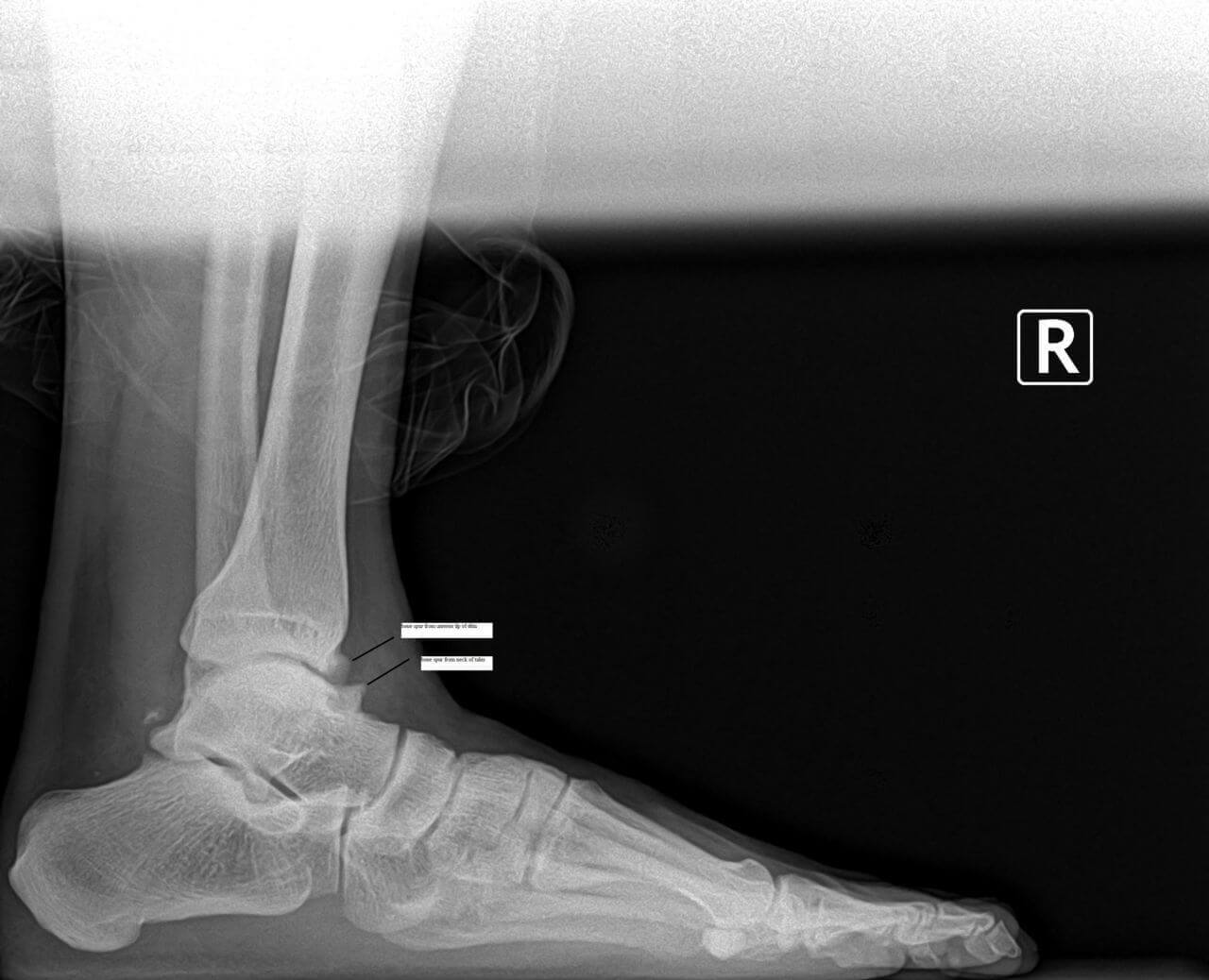
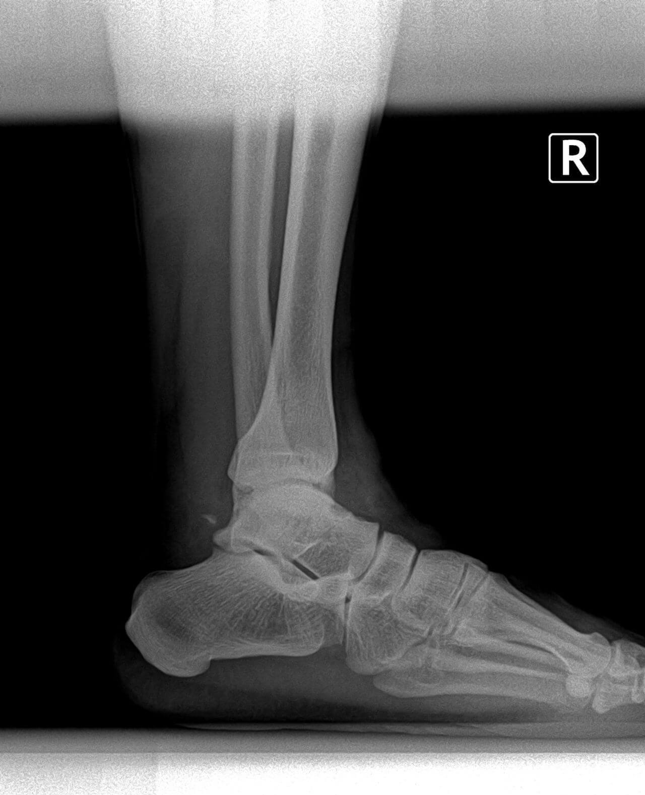
    X-rays help rule out fractures, "fleck fractures" inside the ankle joint, loose bodies, and/or degenerative joint disease (arthritis). Stress X-rays are taken when ligamentous rupture or ankle instability is suspected. When a stress test is taken of your ankle, don't be surprised if the same test is performed on the other ankle. This is done to compare the two ankles, particularly in cases of ligamentous laxity (loose ligaments).

    In the past, more commonly, ankle arthrography has been used. This involves injecting a dye into the ankle joint as it is X-rayed. This helps determine if a rupture of a ligament or tear of the ankle capsule has occurred. However, this procedure does involve some discomfort during the injection process, and, on rare occasions, an allergy to the dye occurs.

    Other diagnostic tests include computerized tomography (CT Scan) to discover injuries of the bone, and magnetic resonance imaging (MRI) to isolate and diagnose specific soft tissue injuries (ligaments, tendons, and capsule). The MRI is very specific, and gives a clear-cut view of these important structures.

    Treatment

    Treatment of an acute ankle injury usually begins with an aggressive physical therapy program that controls early pain and inflammation, protects the ankle joint while in motion, re-strengthens the muscles, and re-educates the sensory receptors to achieve complete functional return to running activity.

    Modalities that decrease pain and control swelling include icing, electrical nerve stimulation, ultrasound, and/or iontophoresis patches. Easy, mild motion, with the limits of pain and swelling, can actually reduce the effects of inflammation. A continued passive motion (CPM) machine can be very helpful in decreasing pain and swelling.

    Resumption of running activity is usually dependent on the runner's limits of pain and motion, and is begun to tolerance. As the runner improves, diagonal running can be prescribed. It is important to protect the runner with braces such as air casts, ankle braces, etc., which help to allow motion at the ankle joint under weight bearing.

    Home exercise programs are very helpful for the post-ankle sprain runner. Proprioception re-education is critical for both the acute as well as the chronic ankle sprain. It may involve using a simple tilt board or more sophisticated proprioceptive training and testing devices.

    For the acute grade III lateral ankle sprain, or complete deltoid tear, complete immobilization is usually recommended for at least four weeks. Afterwards, a removable cast is used to restrict motion and allow for physical therapy. If the ankle does not respond and ankle instability is diagnosed, surgical intervention may be required.

    Today, ankle arthroscopy is a much less invasive procedure than other surgery and allows the ligament to be stabilized with tissue anchors. This eliminates an extended period of immobilization, joint stiffness and muscle atrophy. Post-operatively, this primary ligament repair is protected for approximately a two-to three-week period of time in either a cast or removable cast boot, with daily-continued passive motion, cold therapy, and controlled exercise.

    At three weeks, a simple air cast or ankle brace is applied for an additional three weeks while therapy and rehabilitation is progressing. At six weeks, these devices are used only during running and other athletic activity as a safeguard. As the runner resumes strength and proprioceptive capabilities, the devices are discontinued.

    Conclusion

    When an acute or chronic ankle sprain is not treated, as unfortunately is all too often the case, repeated ankle sprains may occur. Because chronic ankle injuries do not show acute inflammation even when the ankle is weak and unstable, this may set the runner up for another ankle sprain when least suspected. A successive sprain may be more severe than the first, and cause an even more significant injury.

    The most important point to keep in mind when talking about ankle injuries, then, is to prevent the condition from becoming chronic or recurrent.

    So the next time you roll over that stone, or land in that small hole, make sure that your simple ankle sprain is just that: "simple".

    If you don't want to have a swollen ankle all the time while running, don't ignore early warning signs. If you have any doubts about its seriousness, have your podiatrist check your injury.

  • Plantar Fasciitis And Exercise

    The most important thing to remember when exercising with plantar fasciitis is to use pain as your guideline. Runners can be prone to plantar fasciitis due to a developing overuse injury. Once the plantar fasciitis starts, with continued exercise or impact activity, it continues to get worse. By the time it is bad enough that something needs to be done, there is already significant underlying enlargement and swelling of the plantar fascia.

    It is important to remember that plantar fasciitis has both an inflammatory component and mechanical component.  From a mechanical standpoint, it is important to immediately stop doing impact exercise or activities. Here are some tips for relieving pain causing by plantar fasciitis:

    • No impact exercise. Low-impact activities such as aqua jogging and stationary cycling are great alternatives. 
    • Using of a night splint to help stretch out the the calf muscle.
    • Aggressive stretching, but making certain to stretch the calf, not the foot.
    • Taping the foot especially for any limited impact activities or weightbearing activities.
    • Using a Bledsoe plantar fasciitis brace
    • Replacing worn out shoes and wearing shoes with excellent support.
    • For significant cases, the use of prescription orthotics is ideal.
    • Massage therapy and ASTYM physical therapy massage

    From an anti-inflammatory standpoint, the plantar fascia itself is almost always inflamed and this inflammation can be addressed by various approaches:

    • Icing the foot for least 20 minutes directly after physical activity and in the evening
    • An over-the-counter anti-inflammatory can be utilized per label instructions. But, it is important to realize that this can mask the pain and it is possible to then overdo it because one might think that it is healed when it is not.
    • Some forms of physical therapy such as ultrasound therapy or electrical stimulationprovide an anti-inflammatory effect ORIF physically decrease deep swelling (edema).
    • cortisone injection can be beneficial. Ideally, this should be directed at the interface between the plantar fascia and the fat pad and not within the plantar fascia. An injection within the fascia increases the risk of a tear of the fascia.

    More aggressive treatments:

    Exercise guidelines:

    • Use pain as a reference to your allowable activity level.
    • When you are in the acute phase of plantar fasciitis with pronounced pain in the morning and when you get up from sitting, it still too early to return to exercise.
    • Initially, non-weightbearing exercises may be tolerated without an increase in pain that day or the following morning.
    • Use of a recumbent bike, swimming or aqua jogging

    After several weeks (or even a month or longer), the following are good exercise options: elliptical machine, rowing machine, outdoor cycling and spinning class.

    The last phase would be returning to the most pronounced impact activities such as running, basketball or other impact sports. There should be 90-95% improvement with minimal pain before returning to these impact activities.

    Relapse: 
    If you start to increase your activity level, but start having increased pain levels, you have gone too far too fast. It is time to back off very quickly. Remember that some forms of mechanical support will allow you to do more and pay less of a price.

  • The Pros And Cons Of Stretching

    There have always been questions and controversy over whether it is good or bad to stretch. Runners are often concerned about "over-stretching". They worry if they have stretched enough or too much.

    First, why stretch?

    Many coaches and trainers create pre-game or pre-sport warm-ups. They understand that a few minutes spent stretching before an activity can help prevent many common injuries such a sprains, strains and tears. This preparatory activity stimulates circulation to the muscle, connective tissue areas and joints, increasing flexibility and improving range of motion. In short, stretching helps prepare the body for activity.

    Watch a dog as he arises from bed, and lets you know he's ready for exercise. The first movement he engages in is his long stretch. You've seen many other animals practice the same ritual, and yet no one told them to do it! It's a natural experience to prepare the body for quick, propulsive action. Think how you spend your day: sitting at a desk, standing in one position, driving and watching television, sometimes for hours at a time. After maintaining these positions so long, the flexor muscles tighten and shorten, while the extensor muscles require strengthening. Women who wear high heel shoes all day have a similar problem, with calf muscles and Achilles tendons tightening and shortening. Stretching is imperative for them prior to activity.

    After muscles have been stretched to 110% of their resting length, they function much more efficiently. Moving joints help to improve the flow of synovial fluid within the joints. By performing exercises which increase flexibility and strengthen muscles, improved function follows.

    However, stretching has to be done properly. Either the participant or a trainer must be familiar with muscle groups, their origins, and their insertions. Certain exercises are recommended for particular sports, and they usually help to work specific muscle groups.

    The rule of thumb when exercising and stretching is to start distally (towards the ends of the body) and work proximally (towards the core of the body). Muscles to pay attention to are the triceps in the upper body and the hamstrings in the lower extremity. The calf muscles and Achilles tendon need extra attention, since they are usually the first muscles to tighten during the course of the day. Stretching these muscle groups will aid your heel strike, which, in turn, will help with deceleration on the running stride, and allow for a better, more stable, push-off. Other muscle groups that need attention are the adductors (internal rotators) and abductors (external rotators) of the hip. In long-distance running, as well as in in-line skating and cycling, they are very important.

    Since the anterior muscle groups (quadriceps) are usually much more dominant than the posterior (hamstring, gastroc-soleus) muscles, it is imperative to strengthen both muscle groups equally. Otherwise, the outweighing muscle group will cause the weaker group to work harder, and will usually cause a pull or strain. This happens frequently in the latter miles (18-20) of a marathon. Many runners will start to "cramp-up"' an indicator they are overusing weaker muscle groups. As a result, racing times that were excellent midway through the race begin to sour.

    Do’s and Don'ts

    The cardinal rule of proper stretching is: "don't bounce". Ballistic stretching will only cause the muscle receptors to fire, and, rather than stretch, will cause the stretch reflex mechanism to instantly contract.

    The stretch should be performed slowly, and each side should be repeated, holding the stretch for 10 seconds. It is recommended that the stretch be repeated 10 to 15 times.

    Whereas ice is the remedy for muscles and joints after an activity, heat is the prescription for muscles prior to activity. By heating the muscles (warm shower, bath or hot tub) for approximately 10 minutes, muscles will have increased blood supply when they are needed to stretch or fire quickly. If a runner has a pre-existing overuse injury such as tendonitis or muscle strain, the heat will help tremendously. A light massage will also offer beneficial results. This warm-up makes the beginning of the run go smoother, and decreases stiffness at the end or after the run.

    So you're probably asking yourself, can stretching be harmful, or can I overstretch?

    The answer is a definite yes!!!

    Bouncing or overstretching can be counter-productive. It can cause micro-trauma or tears in the muscles or connective tissue. As a result, this can create a weakness that may surface later in the run. Stretching should be performed for a prescribed period of time, generally not to exceed 5 to 10 minutes. Keeping the body in proper alignment when performing the stretch is important to keep in mind. Stretch adequately as many of the muscle groups as will be involved in the activity.

    In the summer months, stretching is not as necessary as it is in the winter. However, it is still recommended as opposed to a slow trot warm-up during the first mile. Running slowly in the beginning of the run in hot climates is often recommended to avoid increased lactic acid build-up and cramps. In cold weather, there is no substitute for good stretching to warm up muscles and prepare them for the run ahead. This help prevents tightness due to the cold, and helps to avoid injury.

  • Anatomy Of The Running Shoe
    As all runners know, running begins with a good foundation. And where do we find that foundation? At the ground level where the rubber meets the road.

    In other words, your shoes, the pieces of leather and rubber that separate your feet from the hard concrete of the road.

    Let's look at the anatomy of a running shoe, and the four sections of the shoe that make it complete.

    The uppers of the shoe may be made of leather or, for the lighter shoes, a synthetic which is lighter, washable and breathable (to reduce heat from the foot). Another component of the upper is the tongue of the shoe, which should be padded in order to cushion the top of the foot against lace pressure. At the back of the shoe, the ankle collar should also be padded to prevent rubbing and irritation of the Achilles tendon.

    The outersole of the shoe is the treaded layer which is glued to the bottom of the midsole. It resists wear, provides traction, and absorbs shock. This is probably the most important layer for the "street fighter" or road runner. The outer sole usually consists of blown rubber, hard carbon rubber, or a combination. The blown rubber is the lightest, but is not durable as pure carbon. The stud or waffle outersoles are excellent for running on soft surfaces such as grass or dirt as they improve traction and stability. On the flip side, the ripple sole is better designed for running on asphalt or concrete surfaces.

    The heel counter is the inflexible material surrounding the heel. It must be made of a material that is both rigid and durable to support and stabilize the heel. Just look at any old shoes, and you will see the wear and breakdown of the inner heel counter, which, over a period of time, tends to lose its stiffness. That's why an external counter is typically placed between the midsole and the base of the heel counter. You will also see a wedge that adds height to the heel and enhances the shoe's ability to absorb shock and reduce strain. The advantage to the added heel height is that it will shorten the Achilles and Gastrocnemius-soleus muscle, reducing the strain upon those important posterior running structures. The downside is that the higher heel height may feel less stable, causing reduced flexibility in the tendon structure.

    The midsole is located between the outersole and the upper. Many regard it as the most important part of the running shoe. It provides cushioning and shock absorption while concomitantly controlling excessive foot motion (pronation/supination).

    The primary materials used in midsoles are ethylene vinyl acetate (EVA) and polyurethane (PU). EVA is a foam that is light and has good to excellent cushioning. The problem is that this material breaks down quickly. In fact, it can break down just sitting in a box in your closet. Compression-molded EVA is one answer, making it harder and more durable. PU is also a foam, usually denser, heavier, and more durable than EVA. PU will stand up longer, but you will give up some of that precious cushioning in return.

    Most shoes today are cushioned with gel, foam, or other manufacturer-specific materials that are designed within the midsole. This type of cushioning will extend the life of the midsole while simultaneously adding increased stability and shock absorption. This typically is where you will see the greatest quality difference between the various companies’ shoes and their models. And this is where the technology wars are being waged.

    Remember, shock absorption is related to how compressible the midsole material can be made. The more the material compresses, the more movement within the shoe is seen. The less the compression of the material, the better the shoe's motion control, but there is a tradeoff in shock absorption. In this case, the shoe may feel harder, and not as soft as the first case scenario.

    Know Your Foot Type

    So how important is it to know what type of foot I have, and how I run?

    Very important! You need to know the basics of running gait and foot types.

    First, when you run, the heel strikes the ground first, usually on the outside (supination). Next, the foot rolls inward and flattens out along the longitudinal arch-pronation. The foot then resupinates by rolling through the ball and rotating outwards. At this point, the foot becomes a rigid lever as it again prepares to push off the ground.

    To find a runner who supinates or pronates just the right amount is rare. Typically, most runners, particularly those who become injured with knee pain, Achilles tendonitis, shin splintsI.T.Band, or heel pain, suffer from either excessive pronation or supination.

    So let's look at those terms again, and how they relate on a runner's gait. Everyone thinks pronation is an evil thing. Wrong! Your foot needs to pronate in order to adapt to uneven surfaces. We all have to pronate to a certain degree. However, excessive pronators whose feet roll inwards too much while running are the runners who develop over-use injuries. The overpronator generally has a flattened type of foot (low arches). You can check this yourself by wetting your feet, and walking on a piece of paper. If you see the whole foot print, including the arch, you can bet you're an overpronator. If you check an old pair of shoes, you'll see a wear pattern to the inside of your shoes, particularly around the big toe.

    Overpronators generally have flexible feet, which creates a very unstable foot. This can lead to many of the overuse injuries previously mentioned. If you are an overpronator, look for a shoe with a lot of motion control, preferably with a board last. A straight-lastedshoe is also recommended for overpronators.

    What about you supinators? The supinator's feet typically roll outward, both in the heel and in the forefoot. You're the ones with the high arches. If you want to see if this is your foot type, go ahead and wet your feet and walk on a piece of paper. If you only see a wet spot of your heel and the ball of the foot, you know you over supinate. When you look at an old pair of shoes, you'll see that they wear excessively on the outside border of the heel, and on the outside of the forefoot near your little toe. You're not in the majority here in this case. Supinators are definitely in the minority compared to pronators.

    The high-arched, supinators' feet are more rigid, and cannot absorb shock as well as an overpronator's feet. Therefore, it stands to reason that with a rigid type of foot the supinator will be subject to more lateral ankle sprains, stress fractures, and pain on the outside of the shin and knee. Supinators should look for a shoe that has better than normal cushioning for added shock absorption, as well as flexibility. Many supinators feel more comfortable with a semi-curved or curved last, due to the shape of their foot.

    Shopping Tips

    So what are some tips for selecting a good running shoe? Both the American Running and Fitness Association and the American Academy of Podiatric Sports Medicine make the following recommendations:

    • Try on both shoes, because your feet may not be the same size and the shoes may not be made symmetrically. Walk and jog around the store for a few minutes; climb stairs, or try jogging on a treadmill if available.
       
    • Try on a couple of different models and sizes so you can make a good comparison. Don't rush your selection.
       
    • The lacing area and tongue should be padded, especially if you have a bony bump (met-cuneiform) on your instep.
       
    • Be sure the sole flexes easily where your foot flexes. Buy shoes with removable insoles so you can modify or replace them with orthotics in needed.
       
    • Allow a half-inch in front of your longest toe when you stand up. Fit shoes in the afternoon or after a workout, when your feet are larger. Feet swell as much as a full size during a good workout.
       
    • The key to finding the best shoe is comfort, not price or brand name. Don't rely on a break-in period;. Shoes should feel good the day you buy them.
       
    • The toe box should allow your toes room to move around. The mid-part of the shoe when laced should hold your foot snugly so that it doesn't slide forward and jam your toes with each step. If the shoe feels tight across your instep, start the laces on the second pair of eyelets.
       
    • The heel counter should fit snugly so your heel won't slip and rub.
       
    • Check the quality control of the shoes. Put them on a flat surface near eye level. The mid-line of the heel counter should be perpendicular to the surface.
       
    • Try on shoes with the socks, inserts, or orthotic devices you plan on wearing.

    It is always suggested that when looking for a good running shoe, first select a good specialty running shoe store with competent salespeople. They are the ones who know the latest in shoe design and performance. Typically, these "fitters" are runners like yourself. They’ve been hired because of their love of running and their interest in their fellow runners. So search them out, and develop a relationship with a store and a person who has been fitting shoes for a period of time. If you have had a history of injury due to a shoe or a biomechanical problem, seek out a podiatrist in your area. He or she will be able to detect what your problem may be, and the right shoe or shoes to look for.

    To extend the life of the shoe, wear them only for running, and let them dry out slowly when wet.

  • Turf Toe

    Activities such as football, basketball, soccer, field hockey and lacrosse show the high incidence of injury to the great toe joint on artificial surfaces. Other non-sporting causes include change in shoe gear, limited range of motion of the great toe joint, and sometimes flat foot conditions.

    Mechanism of Injury

    There are two mechanisms of injury for turf toe. The most common cause is hyperextension of the great toe joint. The great toe joint is hyper-extended as the heel is raised off the ground. An external force is placed on the great toe and the soft tissue structures that support the great toe on the top are torn or ruptured.

    Physical Signs and Symptoms

    Symptoms of acute injury include pain, tenderness and swelling of the great toe joint. Often there is a sudden acute onset of pain during push-off phase of running. Usually, the pain is not enough to keep the athlete from physical activities or finishing a game. This causes further injury to the great toe and will dramatically increase the healing time.

    Injuries to the great toe that cause turf toe are graded into three categories.

    A Grade I turf toe injury is considered to be mild and the supporting soft tissue structure that encompass the great toe are only sprained or stretched. This is the most common type of injury. There is minimal swelling with mild local tenderness and usually no black and blue bruising evident.

    Grade II turf toe injuries are considered moderate in severity. They present with more diffuse tenderness, swelling, restricted range of motion and usually are mildly black and blue in appearance. There is usually a partial tear of the supporting ligaments but no articular cartilage damage.

    Grade III injuries are considered severe in nature because of the considerable swelling, pain on palpation, restriction of range of motion, inability to bear any weight on the injured foot and diffuse black-and-blue appearance of the great toe. There is generally tears to the joint capsule, ruptured, ligaments and possibly compression damage to the articular cartilage of the great toe.

    Treatment

    Treatment is usually centered on an individual basis and the severity of the injury sustained. The following are general principle guidelines for turf toe injuries. The mnemonic "RICE" can be employed. The "R" stands for rest and is the hallmark component to allow for successful healing to occur. However, this is the greatest area of noncompliance because the athlete assumes the injury to be trivial and not severe enough to miss a game or practice. The "I" represents ice, which is usually performed for the first 48-72 hours after the initial injury. Cryotherapy consists of placing the injured toe in a bucket of ice water for 15-20 minute intervals. "C" signifies compression, which is done by taping the great toe in a compression dressing or strapping. The "E" stands for equipment modification or change. For example, the use of a stiffer athletic shoe to resist motion of the great toe or the insertion of an orthotic to increase the support of the great toe.. Additionally, strapping of the great toe to limit motion may allow a highly competitive athlete to return to activities quicker. Non-steroidal anti-inflammatory drugs (NSAID) may be utilized for relief of minor pain as well as to decrease the inflammation of the injury.

    Grade I injuries do well with strapping and usually only require a few days of rest. Grade II injuries should adhere to the "RICE" principles above and usually require one to two weeks of missed practices and games. Grade III injuries are more severe injuries and the healing process may take four to six weeks of recovery time from physical activities. Sometimes, Grade III turf toe injuries do not heal appropriately with conservative care and result in chronic pain and instability. Surgical reconstruction of the joint capsule, ligaments and articular cartilage may be necessary to restore proper alignment and function in these extreme cases.

  • Metatarsal Stress Fracture


    When excessive stress is placed upon the ball of the foot, a hairline break (fracture) of a long metatarsal bone may occur. This occurs most frequently to the second, third, or fourth metatarsal but can occur in any bone. Frequently, the injury is so subtle that you may not recall any specific occurrence. These fractures were at one time referred to as "March Fractures" in soldiers, who developed foot pain after long periods of marching. Stress fractures can occur during sports activities, in overweight individuals, or in those with weakened bones such as osteoporosis.

    Diagnosis

    A typical presentation for someone with a metatarsal stress fracture would be pain and swelling in the ball of the foot, which is most severe in the push off phase of walking. Pressing on the bones in this area of the foot will reproduce the pain. X-rays taken during the first two to three weeks after the injury often will not show any fracture. A bone scan at this stage will be much more sensitive in diagnosing the early stress fracture. The decision to order a bone scan will be up to your doctor. Often times the diagnosis can be made based upon clinical findings, thus making the bone scan unnecessary. After several weeks, an x-ray will show the signs of new bone healing in the area of the stress fracture.

    Treatment

    Treatment for a metatarsal stress fractures usually consists of rest, elevation, and ice initially. Sometimes a compression bandage is applied to help reduce the swelling. Frequently a post-op type of shoe or camwalker is used to prevent you from pushing off the ball of your foot, thus eliminating any additional stress while the bone is healing. Occasionally a short leg walking cast may be applied for a short period of time. Typical healing times range from 4 to 8 weeks. After the fracture is healed, special attention should be paid to using a well-padded insole or a functional orthotic in the shoes to reduce the stress in this area. For those who may have osteoporosis, bone densitometry testing should be done, and appropriate treatment initiated to prevent further weakening of the bones.

5 out of 5 stars
Total Reviews : 232