Information for WA Patients on Common Foot and Ankle Issues | Issaquah Foot & Ankle Specialists

Information is key to treating your foot injury. In our library, we explain the common causes of many foot conditions, including bunions, broken toes, plantar fasciitis, ingrown toenails, and more. Search through our articles to find out more about your condition.
  • Ankle Fusion/Arthrodesis
    What is an ankle arthrodesis?

    The ankle joint consists of portions of three bones: the talar dome, distal tibial plafond and distal fibula. The talus and tibia portions make up over 80% of the articular surface area of the ankle joint. Arthrodesis procedures are the removal of cartilage and any diseased bone from a joint to produce a fusion of at least two bones to create one bone. This removal of cartilage exposes the underlying bone on both sides of the joint. These joints surfaces are then compressed together with some form of fixation to create the fusion. In an ankle arthrodesis, typically the tibial position is fused to the talus. In some cases the fibula is part of arthrodesis, but this is the surgeon’s preference.

    Why is an ankle arthrodesis procedure performed?

    Ankle arthrodesis procedures are typically performed when all conservative options have been attempted and failed. Some of these treatment options, are corticosteroid injections, ankle foot orthoses, anti-inflammatory medications, custom orthotics, ankle braces, and sometimes arthrotomy or arthroscopy. In cases where the pain and/or the deformity is unremitting the arthrodesis is recommended. Some of the conditions that produce unremitting pain and deformity in the ankle joint are:

    1. Post-traumatic arthritis
    2. Rheumatoid arthritis
    3. Infection
    4. Failed ankle implant
    5. Congenital ankle deformities
    6. Neuromuscular disorders (i.e. post-polio syndrome, paraplegia, etc.)
    7. Lateral ankle instability (after failure of operative and non-operative treatment)
    8. Avascular Necrosis (secondary to trauma or metabolic disease states)
    9. Charcot Neuropathy

    Techniques for ankle arthrodesis

    Over the years many techniques have been devised to fuse the ankle joint. Today there are three different techniques that your surgeon may use. They are open technique (use of one or two incisions on the outside and the front of your ankle) with screw fixation, open technique with external fixator fixation and arthroscopic ankle arthrodesis. Most surgeons use all three of these techniques, with the open technique with screw fixation the most commonly used. The technique used is surgeon's preference for your clinical findings, but all have equal fusion rates. Sometimes your doctor may need the use of bone graft, typically taken from your hip area (iliac crest), if you have defects in you ankle from previous trauma or surgery to help support the arthrodesis site.

    What to Expect Following the Procedure

    After most ankle arthrodesis procedures, your surgeon will have you non-weightbearing (no weight on your foot) typically for a period of 6-12 weeks, but this can be extended longer if there is any delay in healing of the arthrodesis site. After the surgery, you will be in a below knee cast or splint to help protect the surgical site and also prevent any movement. After the 6-12 weeks period your surgeon, will typically start you partially weightbearing with your crutches with the use of some type of cast or ankle brace for 4-6 weeks. Once you are fully weightbearing in the cast brace, you will be progressed into high-topped shoe or sneaker. Sometimes modifications have to be made to your shoes, called a rocker-bottomed sole, to aid in push off in ambulation after surgery.

    Some other things that are important to note after the surgery, typically there will be some level of pain or discomfort after the surgery. In a majority of cases, you will be admitted after the surgery to the hospital for pain management. This admission can range from 23 hour to 3-4 days based on an individual’s pain tolerance. When in the hospital and also when you go home it is essential that you keep your foot elevated and use ice as recommended by your surgeon. Ice and elevation will help to reduce the swelling around your foot and ankle that is common after a major surgical reconstruction. The swelling in your foot and ankle can last for 6-9 months and even up to a year. Another thing that is tied closely to the swelling after surgery is a phenomenon that occurs when you get up from after you have had your foot elevated for a period of time. What happens is when you dangle your leg to get up, blood flow will increase into the foot and ankle which will create a throbbing sensation and sometimes a pins and needles sensation in your foot. This is perfectly normal and will go away when you re-elevate your foot.

    Recovery Time

    Time off work depends on the type of ones work as well as the procedures performed. Usually a patient can return to work in 2 to 4 weeks if they are able to work while seated. If a persons job requires standing and walking, return to work may take several weeks when one is able to bear weight. Complete recovery may take six months to a full year.

    Potential Complications

    Complications can occur as with all surgeries, but are minimized by strictly following your surgeons post-operative instructions. The main complications include infection, bone that is slow to heal or does not heal, progression or reoccurrence of deformity, a stiff foot, and the need for further surgery. Many of the above complications can be avoided by only putting weight on the operative foot when allowed by your surgeon.

  • Ankle Fractures
    A fall, an accident, or a hard blow may cause one or more bones in the ankle to fracture or break. Depending on the type of injury, an ankle may fracture in several ways. Spiral fractures may result from twisting injuries. If bone pulls away from bone, the break is often straight. After a bad fall, or a heavy blow, bone may be crushed. Although ankle fractures tend to cause pain, you may be able to walk on the ankle. However, this is not a good idea. Your physician should check any injured ankle.

    Treatment

    Your treatment depends on where and how badly your ankle has been broken. Your physician will feel your ankle and foot for swelling and any displaced bones. He or she may also check to see if the ankle joint still moves. Depending on how the injury occurred, your physician may examine your leg from the knee down. X-rays will be taken to show the type of break, its exact location, and the extent of the damage.

    After a fracture, a cast may be used to hold the bone in its proper position for healing. Sometimes the sections of broken bone must first be realigned. This is called reduction. The type of reduction is based on how far the bone has moved from its normal position.

    Closed Reduction

    If you have a clean break with little soft tissue damage, closed reduction may be used. Before the procedure, you may be given a light anesthetic to relax your muscles. Then your doctor manually readjusts the position of the broken bone.

    Open Reduction

    If you have an open fracture (bone sticking out through the skin), badly misaligned sections of bone, or severe tissue injury, an open reduction will be used. A general anesthetic may be used during the procedure to let you sleep and relax your muscles. Your doctor then makes one or more incisions to realign the bone and repair soft tissues. Screws or plates may be used to hold the bone in place during healing.

    Casting a Fracture

    To make sure the bone is aligned properly, an x-ray is taken. The ankle is then put in a cast to hold the bone in place during healing. You will probably have to wear the cast for 4 to 8 weeks. For less severe fractures, a walking boot, brace, or splint may be all that is needed to hold the bone in place during healing.

    Once your fracture has been treated, your physician will tell you how to help it heal. You may be told to limit your use, take medications, and elevate the foot. If you have a cast, remember to keep it dry. To reduce swelling and control pain, elevate the ankle above the level of your heart. This simple action can help control symptoms throughout your recovery.

    Protect your ankle by giving it enough time to heal. When you do become active again, expect some swelling and stiffness. To build strength and help avoid re-injury, your physician may prescribe physical therapy or home exercise. Ankle support may also help. Exercise increases the flexibility of your ankle. If your physician agrees, try the following exercise. Use your foot to write out the alphabet in the air. Be sure to work from the ankle and foot, not from the knee.

  • Ankle Arthritis

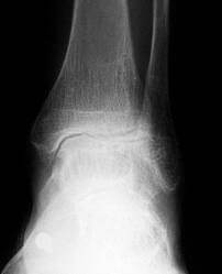
    Ankle arthritis can result from such things as ankle sprains, ankle fractures and infection.  Other conditions that can lead to an arthritic ankle include rheumatoid arthritis or other inflammatory conditions. 

    Ankle arthritis treatment in the Issaquah, WA 98027 area

    Ankle Arthritis results from  a loss of cartilage within the joint that normally provides a lubricated surface for smooth range of motion.  The cartilage begins to wear away and patients often feel a catching, stiffness, swelling or pain with activity.

    Treatment for ankle arthritis can include conservative or surgical care.

    Conservative care:

    • Bracing
    • Injections
    • NSAIDS
    • Physical Therapy

    Surgical care:

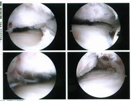
    Ankle Arthroscopy in the Issaquah, WA 98027 area

    • Ankle Arthroscopy
    • Ankle Arthrotomy
    • Ankle Arthrodesis
    • Ankle Joint Replacement
    • Ankle Arthrodiatasis
  • Allergic Reactions, Contact Dermatitis
    There are two types of dermatitis caused by substances coming in contact with the skin: primary irritant dermatitis and allergic contact dermatitis. The primary irritant dermatitis is due to a non-allergic reaction of the skin resulting from exposure to an irritating substance. Allergic contact dermatitis is the allergic sensitization to various substances.

    Primary Irritant Dermatitis

    People who work in areas where their feet are exposed to repeated or prolonged contact to chemicals, oils, or wet cement can develop primary irritant dermatitis. There are certain solutions that people soak their feet in as home remedies. Some of these solutions are safe if used properly, but their improper use can cause a significant contact dermatitis. This can result in skin break down and infection. This is particularly dangerous in people with diabetes; the result can be devastating and limb threatening. A common misconception is the value of soaking in hot water. Some people believe that the hotter the water the better. Quite to the contrary, hot water can cause damage to the skin and result in first or second-degree burns. People will soak their feet in all sorts of solutions. Common solutions are bleach, vinegar, salt water and iodine-based solutions such as betadine. If used properly and under the guidance of a doctor, these solutions can be beneficial. A common mistake that is made is to create solutions that are too strong. Should this occur, irritation to the skin and the development of a rash can develop. The dermatitis that results can also become secondarily infected.

    Allergic Contact Dermatitis

    Allergic contact dermatitis is the result of exposure to substances that sensitize the skin, so that each time one becomes exposed to it again, an inflammatory reaction will occur. Some people are allergic to the substances in the dyes of socks or the materials used to make shoes. The rash that develops is in a pattern that reflects the exposure to the substance. Adhesive tapes can cause an allergic reaction with blisters or a rash developing beneath the tape. Not all blistering or rashes from tape on the feet however are an allergic reaction. Because of the heat and the accumulation of moisture beneath the tape an acute athlete's foot infection may occur.

    Treatment

    Treatment should be directed at the cause of the dermatitis. Burns should not be treated with ointments because ointments are too occlusive and can trap the heat in the burned tissues resulting in further tissue damage. Cool compresses are soothing and can limit the damage caused by the burn. The dermatitis caused by the soaking of the feet in concentrated solutions act like chemical burns and cool compresses are also useful in this instance. Dermatitis caused by an allergic reaction will respond well to topical steroid compounds like hydrocortisone cream. Steroid creams should not be used unless you are certain that the condition is caused by an allergy. Cortisone creams will mask infection and allow infections to get worse while reducing the normal inflammatory reaction associated with infections. Anti-fungal creams are useful in the treatment of athlete's foot that may be caused by occlusion under adhesive tape.

  • Alcoholic Neuropathy

    Peripheral Neuropathy is a nerve condition that affects the arms, hands, legs, and feet. The most common form of peripheral neuropathy is due to diabetes.

    Alcoholic Peripheral Neuropathy

    Alcoholic neuropathy is caused by the prolonged use of alcoholic beverages. Ethanol, the alcoholic component of these beverages, is toxic to nerve tissue. Over time, the nerves in the feet and hands can become damaged resulting in the same loss of sensation as that seen in diabetic neuropathy. The damage to these nerves is permanent. A person with this condition is at the same risk, and should take the same precautions as people with diabetic peripheral neuropathy. Peripheral neuropathy can also be caused by exposure to toxins such as pesticides and heavy metals.

    Treatment For Peripheral Neuropathy

    Treatment for peripheral neuropathy is, for the most part, directed at the symptoms of the condition. Vitamin B12 injections may be helpful if the patient has a vitamin B deficiency. There are certain oral medications that may ease the burning pain that can be prescribed by your doctor. Topical ointments should only be used with the advice of your doctor. Magnetic therapy and Galvanic Stimulation are alternative forms of treatment but results are varied and difficult to quantify.

  • Advice On Shoe Fitting

    For many people, finding a shoe that fits properly can be a frustrating and time-consuming process. Because many people have subtle abnormalities of their feet, the process of finding a proper fitting shoe can be difficult. There are few simple guidelines that if followed can make the process a bit more tolerable. There are several good shoes on the market some of them may even be styles that you may like. With the availability of Internet shopping, the process of finding shoes for the hard-to-fit indiuidual may be made a bit easier.

    When purchasing shoes, it is always a good idea to have the salesperson measure your feet. It is also a good idea to have both feet measured, because in many instances there may be a difference in the size of your feet. If you have two feet that are not the same size, it is recommended that you buy shoes to fit the bigger foot. Our feet change just like our eyes do as we get older. A person's feet tend to become a bit longer and wider. Women, during pregnancy have a tendency for their shoe size to change. This is because during pregnancy a woman's body produces a hormone called elastin. This hormone softens the ligaments about the pelvis to assist during delivery. Unfortunately, the hormone also affects other ligaments in the body. The ligaments in the foot are particularly affected. This coupled with an increase in weight and a change in the center of gravity causes many women to experience a change in their shoe size. Our feet also have a tendency to change size during the course of the day. Shoes that may feel comfortable in the morning may feel tight and uncomfortable later in the day. This occurs because of a variable amount of swelling in the feet that occurs as the day goes on. Therefore it is a good idea to buy your shoes later in the day.

    The shape of the shoe is important, but surprisingly little attention is paid to this feature of the shoes we buy. The "Last" of the shoe determines the over all shape of the shoe. The shoe "last" may be straight or curved. To determine the "last" of the shoe, turn the shoe upside down and look at the sole. Imagine a line that goes through the center of the heel of the shoe and then out the center of the toe area of the shoe. You might be surprised what you find. In "curve lasted shoes" this imaginary line is in the shape of a curve, usually curving inward. Many shoemakers make curve lasted shoes. This is particularly true in sports shoes. A "straight lasted shoe" will have this imaginary line almost straight from the heel to the toes. Now, look at the shape of your foot. Have you ever wondered why your shoes seemed to wear out in the upper part about the toe box in such a funny way? The reason may very well be that you have a rather straight foot and you are wearing a curve shaped shoe. Curve lasted shoes can aggravate a number of foot problems. These shoes can cause an excessive amount of pressure on the outside of the foot. This has the potential of irritating existing problems like bones spurs in the fifth toe, soft corns between the fourth and fifth toe, and tailors bunions. Another area of the foot that can become irritated is along the outside of the foot called the "styloid process". The styloid process is the base of the long bone (metatarsal) behind the fifth toe. In some people the styloid process is more prominent and subject to irritation by shoe pressure. There is also a powerful tendon that attaches into the foot in this area from a muscle on the outside of the lower leg. This tendon and some other small tendons on the top of the foot can be irritated by the curve lasted shoe.

    Shoe manufactures make curved lasted shoes because they believe that by curving the foot inward it causes greater stability to the foot. In theory they are correct but shoes rarely are made of materials that are strong enough to influence foot function. Instead, as a person wears the shoe, the shoe over time becomes mis-shapen and can cause irritation to areas on the outside of the foot. The shoe manufactures have been a bit more successful at producing "motion control" shoe wear in sports shoes, but even there the shoe will rarely be able to hold up to the deforming forces of the foot over time. If a person has an abnormality of their foot that requires some degree of "motion control" they are better advised to seek the advice of a foot specialist who can determine their needs and prescribe a device that corrects abnormal function of the foot. These devices called orthotics fit into normal shoes and last for several years. In many cases the use of an orthotic will correct abnormal wear patterns seen in a persons shoes.

    When selecting a good sports shoe there are a few simple guidelines to follow. First of all fit the shoe to the shape of your foot. In other words, if you have a fairly straight looking foot choose a shoe that has a straighter last to it. Secondly, consider sport shoes that are relatively rigid in the heel portion of the shoe. Heel stability is important in almost all cases. Additionally, look for a shoe that is fairly flexible in the forefoot area. If the shoes does not easily flex in the forefoot then as the heel comes off the ground during walking and running the big toe is unable to flex properly. Adequate movement of the big toe joint is important for normal foot function. There are two more things to check before you purchase the shoe. Place the shoe on a firm flat surface and observe what the back of the heel of the shoe looks like. The heel of the shoe should be relatively perpendicular to the surface the shoe is sitting on. If the back of the shoe is angled in one direction or another this could indicate a defect in the manufacture of the shoe. Lastly, put you hand inside the shoe and check for any defects in the seams of the shoe. Seams that are prominent have the potential to cause irritation to areas on the foot.

    Diabetic patients need to be particularly aware of the type of shoes that they wear. This is especially true if they have poor circulation, numbness or a loss of sensations in their feet (neuropathy). Shoes should be purchased that have adequate room in the toe box area. The upper of the shoe should be of soft leather with few or no seams. Extra depth shoes are available that meet the needs of many diabetic patients. In many instances Medicare will reimburse for one pair of shoes per year if the patients doctor recommends extra depth or special shoes. There must be adequate documentation in the doctor's medical record for the need for the shoes. Mnay foot doctors offer diabetic shoes as part of their practice.

  • Actinic Keratosis
    A cancer causing lesion that can occur on the feet are called Actinic Keratosis. Although most commonly found in sun-exposed areas of the body such as the face, ears, and back of the hands, these lesion can also occur on the foot. They are characterized as either flat or elevated with a scaly surface. They can either be reddish or skin colored. On the foot they are frequently mistaken for planters warts. These lesions are the precursor of epidermis carcinoma. Treatment for these lesions should be through as they are definitely precancerious. Treatment consists of freezing the lesions with liquid nitrogen or sharp excision.
  • Acrocyanosis

    Acrocyanosis is a vasospastic disorder affecting the arteries supplying blood to the skin of the hands and feet. Vasospasm refers to the arteries going into spasm and blocking the flow of blood. These small arteries carry oxygen and nutrients through the blood to the skin of the hands and feet. When the blood cannot flow through, the skin will lack the necessary oxygen required, and turn a dark blue to purple color. This characteristic color is called cyanosis, hence the name acrocyanosis. It is not a common condition. It is seen more frequently in woman than in men.

    Unlike the vasospasm seen in Raynaud's disease which may last several minutes to several hours, the vasospasm in acrocyanosis is more persistent. In addition, the vasospasm in Raynaud's disease affects the small arteries supplying blood to the fingers and toes. In acrocyanosis the vasospasm affects the arteries supplying blood flow to the skin of the hands and/or feet. Therefore the skin damage and ulcerations seen in Raynaud's disease are not present in acrocyanosis. Lastly, Raynaud's disease goes through a typical triphasic or biphasic color change. In contrast, acrocyanosis maintains its characteristic blue skin coloration.

    Diagnosis

    Typical symptoms and signs of acrocyanosis of the hands or feet, are a persistently cold temperature and blue discoloration. They often feel sweaty or moist, and swelling may be present. The blue cyanosis usually appears worse upon exposure to cold, and improves upon warming. Rarely is there any pain associated. Normal arterial pulses are always present in the hands and feet since there is no blockage of the larger arteries of the arms or legs.

    Treatment

    Generally the treatment is a common sense approach to preventing cold exposure and keeping the feet dry. This may involve the use of insulated boots, thin polypropylene liner socks to wick the moisture away from the skin, and an insulated sock to maintain normal skin temperature. Generally no other treatment is necessary. Vasodilators have been tried with limited success. In extreme cases a surgical procedure called a sympathectomy has been performed to relax the persistent vasospasm. This surgery is rarely necessary, and seldom recommended.

  • Surgical Treatment Of Adult Aquired Flatfoot
    Adult acquired flatfoot deformity or posterior tibial tendon dysfunction is a gradual but progressive loss of ones arch. The posterior tibial muscle is a deep muscle in the back of the calf. It has a long tendon that extends from above the ankle and attaches into several sites around the arch of the foot. The muscle acts like a stirrup on the inside of the foot to help support the arch. The posterior tibial muscle stabilizes the arch and creates a rigid platform for walking and running. If the posterior tibial tendon becomes damaged or tears the arch loses its stability and as a result, collapses causing a flatfoot. Adult flatfoot deformity can occur in people of all ages and gender however, it occurs most commonly in sedentary middle aged to elderly females. There are several risk factors for posterior tibial tendon dysfunction that include: obesity, steroid use, systemic inflammatory diseases such as rheumatoid arthritis, trauma, being born with a low arch, and diabetes. It occurs most commonly in one foot however, it can occur in both feet especially in people with systemic diseases such as diabetes and rheumatoid arthritis.

    There are four stages of posterior tibial tendon dysfunction. In the first stage the posterior tibial tendon is inflamed but has normal strength. There is little to no change in the arch of the foot. In stage two the tendon is partially torn or shows degenerative changes and as a result loses strength. There is considerable flattening of the arch without arthritic changes in the foot. Stage three results when the posterior tibial tendon is torn and not functioning. As a result the arch is completely collapsed with arthritic changes in the foot. Stage four is identical to stage three except that the ankle joint also becomes arthritic.

    Surgical Treatment

    Surgical correction is dependent on the severity of symptoms and the stage of deformity. The goals of surgery are to create a more functional and stable foot. There are multiple procedures available to the surgeon and it may take several to correct a flatfoot deformity.

    Stage one deformities usually respond to conservative or non-surgical therapy such as anti-inflammatory medication, casting, functional orthotics or a foot ankle orthosis called a Richie Brace. If these modalities are unsuccessful surgery is warranted. Usually surgical treatment begins with removal of inflammatory tissue and repair of the posterior tibial tendon. A tendon transfer is performed if the posterior tibial muscle is weak or the tendon is badly damaged. The most commonly used tendon is the flexor digitorum longus tendon. This tendon flexes or moves the lesser toes downward. The flexor digitorum longus tendon is utilized due to its close proximity to the posterior tibial tendon and because there are minimal side effects with its loss. The remainder of the tendon is sutured to the flexor hallucis longus tendon that flexes the big toe so that little function is loss.

    Stage two deformities are less responsive to conservative therapies that can be effective in mild deformities. Therefore, these patients may be candidates for a 15 minute outpatient procedure to correct the flexible flatfoot deformity which is referred to as hyperpronation. The procedure is called a Subtalar Arthroereisis. It involves the placement of an implant in the space under the ankle joint (sinus tarsi) to prevent only the abnormal motion, but still allowing normal motion. This brief procedure only requires very little recovery time, and is completely reversible, if necessary. Your surgeon can consult you about this exciting, life-changing procedure, or more information can be obtained at www.hyperpronation.com.

    Stage three deformities are better treated with surgical correction, in healthy patients. Patients that are unable to tolerate surgery or the prolonged healing period are better served with either arch supports known as orthotics or bracing such as the Richie Brace. Surgical correction at this stage usually requires fusion procedures such as a triple or double arthrodesis. This involves fusing the two or three major bones in the back of the foot together with screws or pins. The most common joints fused together are the subtalar joint, talonavicular joint, and the calcaneocuboid joint. By fusing the bones together the surgeon is able to correct structural deformity and alleviate arthritic pain. Tendon transfer procedures are usually not beneficial at this stage.

    Stage four deformities are treated similarly but with the addition of fusing the ankle joint.

    Recovery Time

    Time off work depends on the type of work, as well as the surgical procedures performed.

    Potential Complications

    Complications can occur as with all surgeries, but are minimized by strictly following your surgeon?s post-operative instructions. The main complications include infection, bone that is slow to heal or does not heal, progression or reoccurrence of deformity, a stiff foot, and the need for further surgery. Many of the above complications can be avoided by only putting weight on the operative foot when allowed by your surgeon.

  • Surgical Removal Of Giant Cell Tumors

    This tumor was once thought to be a cancer of a tendon sheath. It is now known to be a benign non-cancerous tumor of a tendon sheath. These masses are generally found on the toes, top of the foot or sides of the foot. They are always closely associated with a tendon sheath. They can also occur deep inside the foot. They slowly enlarge but never grow any larger than 4cm in size. They are firm irregular masses that are commonly painful. The pain seems to be a result of the tumor pressing firmly on the surrounding tissues and due to the interference with the function of the tendon the mass is growing from. As the tendon grows it can press so firmly on the bone it lays next to, that it can cause erosion of the bone. It is because of this erosion of bone that the tumor was once thought to be cancerous. Cancerous tumors can have the characteristic of invading bone through aggressive and destructive means. The erosion of the bone associated with giant cell tumors is due to pressure on the bone and not due to the invasion of the bone by the tumor. Other common soft tissues masses that may occur in the foot are ganglions, fibromas.

    Diagnosis

    The diagnosis of a giant cell tumor is generally made by a pathologist following removal of the mass. Clinical history of the mass may give the surgeon an idea of what they might expect when removing the mass. X-rays may show the shadow of the mass, and in 10-20% of the cases, may demonstrate bone erosion. The mass is firm and nodular, and always connected to a tendon. A MRI may be useful in determining the extent or size of the mass.

    Treatment

    Treatment of giant cell tumors is the excision of the tumor. Some physicians may attempt to inject the mass with cortisone in an attempt to shrink the mass.

    The Procedure

    The surgical excision of giant cell tumors is generally preformed in an out patient surgery center. Depending on the location of the mass the surgery may be preformed under a local anesthesia, with intravenous sedation or general anesthesia. Following administration of the anesthesia an incision is placed over the mass. The mass is then carefully dissected free from the surrounding soft tissues. Following the closure of the surgical site a gauze compressive dressing is applied. Depending upon the location of the mass the surgeon may apply a splint or below the knee cast. In some instances the surgeon may prefer that the patient use crutches for a few days or for as long as three weeks.

    Recovery Period

    The recovery period depends upon the location of the mass and the extent of the soft tissue dissection necessary to remove the mass. The sutures are left in place for 10 – 14 days. During this period of time the patient should limit their activities and keep the foot elevated above their heart. It is also important to keep the bandage in place and keep the surgical site dry. If the patient has been instructed to wear a removable cast or use crutches it is important that they follow the surgeons instructions. Time off from work will depend upon the level of activity required of the job and the shoes necessary for work. Generally a minimum of one week off from work is necessary. If the patient can return to work while wearing a cast and they are allowed to perform light duty they may be able to return to work after one week.

    Possible Complications

    The surgery is generally successful and without complications. However, as with any surgical procedure there are potential complications. Possible complications include, infection, excessive swelling, delays in healing, tendon or nerve injury. Because the mass is a growth from a tendon, removal of the mass may require the excision of a portion of healthy tendon. This can weaken the tendon or cause scaring of the tendon. Additionally there may be small skin nerves in the area of the tumor that may have to be sacrificed when removing the mass. If this occurs there may be small areas of patchy numbness on the skin following the procedure. This is generally not a significant problem. On occasion a nerve may get bound down in scar tissue and cause pain following the surgery. Recurrence of the mass is also possible but generally not considered a complication of the procedure.

5 out of 5 stars
Total Reviews : 232