Information for WA Patients on Common Foot and Ankle Issues | Issaquah Foot & Ankle Specialists

Information is key to treating your foot injury. In our library, we explain the common causes of many foot conditions, including bunions, broken toes, plantar fasciitis, ingrown toenails, and more. Search through our articles to find out more about your condition.
  • Common Digital Deformities

    Deformities of the toes are common in the pediatric population. Generally they are congenital in nature with both or one of the parents having the same or similar condition. Many of these deformities are present at birth and can become worse with time. Rarely do children outgrow these deformities although rare instances of spontaneous resolution of some deformities have been reported.

    Malformation of the toes in infancy and early childhood are rarely symptomatic. The complaints of parents are more cosmetic in nature. However, as the child matures these deformities progress from a flexible deformity to a rigid deformity and become progressively symptomatic. Many of these deformities are unresponsive to conservative treatment. Common digital deformities are underlapping toes, overlapping toes, flexed or contracted toes and mallet toes. Quite often a prolonged course of digital splitting and exercises may be recommended but generally with minimal gain. As the deformity becomes more rigid surgery will most likely be required if correction of the deformity is the goal.

    Underlapping Toes

    Description

    Underlapping toes are commonly seen in the adult and pediatric population. The toes most often involved are the fourth and fifth toes. A special form of underlaping toes is calledclinodactyly or congenital curly toes. Clinodactyly is fairly common and follows a familial pattern. One or more toes may be involved with toes three, four, and five of both feet being most commonly affected.

    The exact cause of the deformity is unclear. A possible etiology is an imbalance in muscle strength of the small muscles of the foot. This is aggravated by a subtle abnormality in the orientation on the joints in the foot just below the ankle joint called the subtalar joint. This results in an abnormal pull of the ligaments in the toes causing them to curl. With weight bearing the deformity is increased and a folding or curling of the toes results in the formation of callus on the outside margin of the end of the toe. Tight fitting shoes can aggravate the condition.

    Treatment

    The age of the patient, degree of the deformity and symptoms determine treatment. If symptoms are minimal, a wait and see approach is often the best bet. When treatment is indicated the degree of deformity determines the level of correction. When the deformity is flexible in nature a simple release of the tendon in the bottom of the toe will allow for straightening of the toe. If the deformity is rigid in nature then removal of a small portion of the bone in the toe may be necessary. Both of these procedures are common in the adult patient for the correction of hammertoe deformity. If skin contracture is present a derotational skin plasy may be required.

    Overlapping Toes - Overlapping Fifth Toe

    Description

    This deformity is characterized by one toe lying on top of an adjacent toe. The most common toe involved is the fifth toe. When one of the central toes is involved the second toe is most commonly affected. The etiology of the condition is not well understood. It is though that it may be caused by the position of the fetus in the womb during development. The condition my run in families so there may be a hereditary component to the deformity.

    Treatment

    Effective conservative treatment depends upon how early the diagnosis is made. In infancy, passive stretching and adhesive tapping is most commonly used. This may require 6 to 12 weeks to accomplish and reoccurrence is not uncommon. Rarely will the deformity correct itself. As the individual matures the deformity becomes fixed. When surgical correction is warranted a skin plasty is required to release the contracture of the skin associated with the deformity. Additionally a tendon release and a release of the soft tissues about the joint at the base of the fifth toe may be required. In severe cases the toe may require the placement of a pin to hold the toe in a straightened position. The pin, which exits the tip of the toe, may be left in place for up to three weeks. During this period of time the patient must curtail their activities significantly and wear either a post-operative type shoe or a removable cast. Excessive movement at the surgical site can result in a less than desirable result. The pin can be easily removed in the doctor's office with minimal discomfort. Following removal of the pin splinting of the toe may be required for an additional two to three weeks.

    Hammertoes and Mallet Toes

    Description

    Another common digital deformity is contracture of the toes in the formation of hammertoes and mallet toes. Hammertoes are described in depth in another article. Mallet toes are a result of contracture of the last joint in the toe. In the pediatric population it is often flexible and not painful. Over time the deformity becomes rigid and a callus may form on the skin overlying the joint at the end of the toe. Additionally the toenail may become thickened and deformed form the repetitive jamming of the toe while walking. The deformity usually involves one or two toes, with the second toe most commonly affected. Mallet toes have several etiologies. Longer toes that are forced against a short toe box in the shoe will, over time, develop a contracture of the last joint in the toe causing a mallet toe.

    Treatment

    Conservative treatment consists of padding and strapping the toes into a corrected position. This treatment may alleviate the symptoms but will not correct the deformity. Diabetic patients often develop ulcerations on the ends of their toes secondary to mallet toe deformity and the pressure that results from the toe jamming into the shoe. When standing, the toe will demonstrate a contracture, with the tip of the toe facing downward into the floor. If the deformity is flexible a simple release of the tendon in the bottom of the toe will allow straightening of the toe. Following the procedure the patient must avoid shoes that cause jamming of the toe or the deformity can reoccur. When the deformity is rigid surgical correction requires the removal of a small section of bone in the last joint of the toe. On occasion fusion of the last two bones in the toe may be necessary. This requires removing the cartilage from the last joint in the toe and pinning the bones together. When the bone heals it forms a single bone and the toe remains in a straightened position. Healing time is dependent upon the procedure selected. If a tendon release is performed the patient my return to a roomy shoe within a week. If the toe is straightened by removing a section of the bone in the toe it make ten days to three weeks for a patient to return to normal shoes. If a fusion is performed to straighten the toe, the patient may not return to normal shoes for 6 to 8 weeks. Time off from work will depend upon the type of shoe gear that must be worn and the level of activity necessary to perform the job. A minimum of three to four days off from work is generally recommended and longer if the job responsibilities can not be modified to accommodate the normal healing time for the surgery.

  • Cocked Up Big Toe - Hallux Hammertoe

    The big toe, called the Hallux, is made up of two small bones called phalanges. This condition presents as a cocking up of the big toe at the joint between these two small bones. In the early stages of the condition the deformity is flexible, in later stages the deformity becomes rigid. It is caused by a variety conditions. Neurological diseases that cause muscle weakness or muscle imbalance in the muscles of the lower leg can result in the formation of Hallux hammertoe. This is commonly seen in patients after they have suffered a stroke or Cerebral Vascular Accident. Damage to certain areas of the brain during a stroke will frequently result in weakness and/or paralysis on one side of the body. If the stroke is not severe the patient may recover a majority of the function of the muscles in the legs and feet. However a residual result may be a cocking up of the big toe.

    Other causes of the condition include damage or laceration to the tendon on the bottom of the big toe. Surgery to correct bunion deformities, in rare cases, may result in an imbalance of the structures about the big toe joint and cause the condition. An additional cause of hallux hammertoe is the absence of two small bones, called sesamoid bones, which are normally present beneath the big toe joint. There is an uncommon condition where a person may be born without these bones. More commonly however, the absence of one or both of the sesamoid bones is due to their surgical removal. In the course of correcting a bunion deformity one of the sesamoid bones may be removed. In another situation, a fracture of one or both of the sesamoid bones may result in the necessity to remove them to cure the pain associated with the injury (See the description of sesamoditis).

    A high arched foot may also result in the formation of, not only a hallux hammertoe, but also hammertoes of all of the toes.

    A consequence of having a hallux hammertoe is irritation on the top of the toe from shoe pressure or the development of a painful callus on the end of the big toe. People who have had a stroke or who have diabetes with peripheral neuropathy may not have pain associated with the callus on the end of the toe. These areas may ulcerate and become infected.

    Diagnosis

    The diagnosis of hallux hammertoe is made by clinical exam. An x-ray is useful in determining the degree of the deformity and the condition of the joint. The presence or absence of the sesamoid bones is also made using an x-ray. If a neurological condition has not been identified and there is absence of trauma or previous surgery in the area, then evaluation by a neurologist may be appropriate.

    Treatment

    The need for treatment is based upon the level of symptoms the patient may be experiencing. Splitting the toe in an attempt to straighten it is of little value and is certain to fail. If treatment is needed, surgical correction of the deformity has the greatest level of success.

    If the deformity is flexible a simple tendon release procedure can be performed. This consists of making a small incision on the side of the toe and cutting the tendon in the bottom of the toe. If an ulceration or open sore is present on the end of the toe cutting the tendon to relax the toe may be all that is necessary to allow the ulceration to heal. This procedure can easily be performed in the doctor's office under a local anesthesia. Following the surgery a dressing is applied to splint the toe in a straighten position. The sutures and the bandage are kept in place for 7 to 10 days. The patient should keep their activities to a minimum during this period of time and keep the area dry. A post-operative type of shoe is worn to accommodate the bandage. Generally a patient can return to normal shoes within two weeks and resume complete normal activities in three weeks.

    If the deformity is rigid then fusion of the joint will be necessary to correct the deformity. Under certain circumstances the foot surgeon may elect to fuse the toe when the deformity is flexible. Fusion of the toe requires removing bone at the level of the joint in the toe. The articular surfaces of the joint are removed and the two small bones are abutted up against one another and held in place by a small screw. This fuses the two pieces of bone together resulting in permeate straightening of the toe. This procedure is generally performed in an out-patient surgery center or hospital. The surgery can be performed under a local anesthesia but the patient and surgeon may prefer to use a twilight anesthesia for the patient's comfort. Following the surgery a fluff dressing is applied. The sutures will remain in place for 7 to 10 days. During this period of time the patient should significantly reduce their activities and keep their foot elevated. It takes 6 weeks or longer for the bones to fuse. During this period of time the patient should wear a stiffed-soled post-operative type of shoe. Bending of the toe will delay or inhibit the fusion of the bones. Quite often it takes three months before the patient can return to full-unrestricted activity.

    Possible Complications

    Possible complications include infection, excessive swelling, and delays in healing or failure of the bones to fuse. Overall the procedure has a very high success rate. On occasion, over time, the screw may begin the cause irritation on the tip of the toe and have to be removed.

  • Chronic Ankle Instability

    What Is Chronic Ankle Instability?
    Chronic ankle instability is a condition characterized by a recurring “giving way” of the outer (lateral) side of the ankle. This condition often develops after repeated ankle sprains. Usually the “giving way” occurs while walking or doing other activities, but it can also happen when you’re just standing. Many athletes, as well as others, suffer from chronic ankle instability.

    People with chronic ankle instability often complain of:

    • A repeated turning of the ankle, especially on uneven surfaces or when participating in sports
    • Persistent (chronic) discomfort and swelling
    • Pain or tenderness
    • The ankle feeling wobbly or unstable

    Causes
    Chronic ankle instability usually develops following an ankle sprain that has not adequately healed or was not rehabilitated completely. When you sprain your ankle, the connective tissues (ligaments) are stretched or torn. The ability to balance is often affected. Proper rehabilitation is needed to strengthen the muscles around the ankle and “retrain” the tissues within the ankle that affect balance. Failure to do so may result in repeated ankle sprains.

    Repeated ankle sprains often cause – and perpetuate – chronic ankle instability. Each subsequent sprain leads to further weakening (or stretching) of the ligaments, resulting in greater instability and the likelihood of developing additional problems in the ankle.

    Diagnosis
    In evaluating and diagnosing your condition, the foot and ankle surgeon will ask you about any previous ankle injuries and instability. Then he or she will examine your ankle to check for tender areas, signs of swelling, and instability of your ankle as shown in the illustration. X-rays or other imaging studies may be helpful in further evaluating the ankle.

    Non-Surgical Treatment
    Treatment for chronic ankle instability is based on the results of the examination and tests, as well as on the patient’s level of activity. Non-surgical treatment may include:

    • Physical therapy. Physical therapy involves various treatments and exercises to strengthen the ankle, improve balance and range of motion, and retrain your muscles. As you progress through rehabilitation, you may also receive training that relates specifically to your activities or sport.
    • Bracing. Some patients wear an ankle brace to gain support for the ankle and keep the ankle from turning. Bracing also helps prevent additional ankle sprains.
    • Orthotics.  Custom molded prescription orthotic devices may be prescribed.
    • Medications. Nonsteroidal anti-inflammatory drugs (NSAIDs), such as ibuprofen, may be prescribed to reduce pain and inflammation.

    When Is Surgery Needed?
    In some cases, the foot and ankle surgeon will recommend surgery based on the degree of instability or lack of response to non-surgical approaches. Surgery usually involves repair or reconstruction of the damaged ligament(s). The surgeon will select the surgical procedure best suited for your case based on the severity of the instability and your activity level. The length of the recovery period will vary, depending on the procedure or procedures performed.

  • Chemical Neurolysis For The Treatment Of Neuromas
    The chemical destruction of the nerve, called neurolysis, is an older form of treatment that has recently come back into vogue. This treatment requires a series of injections of ethanol mixed with a local anesthetic. The injections are given into the area of the neuroma. Nerve tissue has a natural affinity for ethanol, and it is readily absorbed into the nerve. Ethanol, however, is toxic to nerve tissue and with repeated exposure, will destroy the nerve. The rate of success is variable, but has been reported to be over 60%. Many insurance plans will not pay for weekly injections and require the doctor to wait a minimum of ten days between injections before they will reimburse for the procedure. This likely reduces the rate of success for this treatment, because during the time between the injections, the nerve will attempt to repair itself. One way to solve this delay is for the patient to pay for those injections not paid for by the insurance plan. The disadvantages for this form of treatment are the need for repeated visits to the doctor’s office, and the occasional pain in the area of the injection the following day or two after it has been administered. The advantages to this form of treatment is that it requires a minimal amount of time off of work and the overall cost as compared to the surgical removal of the nerve. If this form of treatment fails, then surgical removal is the only option that remains.
  • Charcot Joint Disease (Neuroarthropathy)

    Charcot joint disease was given its name by the French neurologist Jean-Marie Charcot in 1868. He noted a bizarre pattern of bone destruction in patients with tertiary syphilis and absent sensation. In 1936, William Jordan described a similar pattern in patients with diabetes.

    Theories as to the cause of the Charcot joint disease abound. However, certain predisposing factors appear to be necessary in order for Charcot to develop. First, peripheral sensory neuropathy or total absence of sensation must be present. Second, circulation is most commonly normal. Third, there is often a history of a preceding injury. This is often so minimal the patient is unable to recall any injury or trauma.

    Who Gets Charcot?

    Charcot joint disease was originally described in patients with tertiary syphilis and absent sensation. However, once penicillin was discovered, the incidence of syphilis dropped dramatically. In general, any disease process that results in loss of sensation in the lower extremity can lead to Charcot joint disease. Today, the leading cause of Charcot joint disease is diabetes mellitus. It is estimated that 1 out of 700 patients with diabetes will develop Charcot joint. Other entities, which can lead to Charcot joint, include: chronic alcoholism, leprosy, hereditary insensitivity to pain, syringomyelia and multiple sclerosis.

    A second key component to the development of Charcot is the history of a preceding injury to trauma to the foot or ankle. Often this injury goes undetected because of a lack of normal sensation. Additionally, because of poor sensation, the injury may not be perceived to be serious. Consequently, the patient will continue with normal or near normal activity leading to further fracture and dislocation of the involved joints and bones.

    Patients who develop Charcot joint will also have good to excellent circulation to their feet. It is uncommon for patients with significant peripheral vascular disease to develop Charcot joint disease.

    What are the Symptoms?

    Patients who develop Charcot joint disease most commonly notice unexplained swelling in their foot or ankle. Because of underlying loss of sensation, this swelling is often painless. On occasion, there may be redness localized to the top of the foot or ankle. There will also be increased warmth to the foot, indicating localized inflammation. Rarely will there be bruising unless the injury is significant.

    Because Charcot joint disease shows swelling and redness, it is often misdiagnosed. The most common misdiagnoses include: cellulitis, osteomyelitis, tendonitis and gout. Failure to diagnosis and treat this entity early and appropriately can lead to foot and ankle deformity.

    Diagnosing Charcot Joint Disease

    The diagnosis is primarily based on the clinical examination. A diagnosis of Charcot joint must be considered in patients with diabetes who present with a warm, swollen foot or ankle without pain. Another critical feature in the diagnosis of Charcot joint is the presence of crepitus or "grinding" in the involved joints. This represents the actual unstable bone fragments moving against each other.

    X-rays at this stage will often confirm the diagnosis, as these will often show fragmentation of bone and disruption of joints. Occasionally, if the x-rays fail to show any disruption and a diagnosis of Charcot joint disease is still being considered, bone scans can be helpful.

    Special studies such as CT scans or MRI are rarely necessary to make the diagnosis. A CT scan can be useful if reconstructive surgery is planned. An MRI can be helpful if an infection with abscess formation is suspected.

    Treatment

    Early diagnosis and treatment is critical to a successful outcome. Once the diagnosis of Charcot joint disease is made, initial treatment should consist of total non-weightbearing and immobilization of the involved extremity. This often requires the use of crutches or a walker. Additionally, a removable cast or brace to protect and immobilize the foot and ankle may be necessary. The duration of non-weightbearing and immobilization will depend on the joints affected and the degree of destruction. As a rule, the larger the joint, the longer the duration of non-weightbearing needs to be.

    Serial x-rays and resolution of the clinical features of the disease determine the healing of the involved joints. Clinically, one would expect to see reduction of swelling, decrease in skin temperature and decrease in joint crepitus. On x-rays, one would expect to see resorption of bone debris and fragments with deposition of new bone and fracture healing. Protected weightbearing may be initiated when the clinical features of the disease show improvement, especially loss of joint crepitus. Weightbearing can be increased gradually as long as there is no re-exacerbation of the disease.

    When foot and ankle deformity develops, custom orthoses and special shoes may be necessary to prevent foot ulcerations and provide stability during ambulation. When ulcerations develop and resist conservative treatment, surgery may be necessary to prevent loss of the foot. Additionally, if there is severe instability, reconstructive surgery of the foot and ankle may be necessary to provide a stable platform for ambulation and to avoid lower leg amputation.

    Complications

    The most common complication of Charcot joint disease is foot and ankle deformity. This can occur even following early and appropriate treatment. This typically results from significant bone and joint destruction that the functional integrity of the foot is sacrificed. Chronic ulcerations may occur as a result of these deformities. Surgery may be necessary to prevent limb loss. Another complication that may occur is instability of the foot and ankle during ambulation. This once again is related to the joints affected and the degree of bone destruction. Severe instability can lead to lower leg amputation. Major reconstructive surgery may be necessary to prevent this complication of Charcot joint disease.

  • Charcot Foot

    What Is Charcot Foot?
    Charcot foot is a condition causing weakening of the bones in the foot that can occur in people who have significant nerve damage (neuropathy). The bones are weakened enough to fracture, and with continued walking the foot eventually changes shape. As the disorder progresses, the joints collapse and the foot takes on an abnormal shape, such as a rocker-bottom appearance.

    Charcot foot is a very serious condition that can lead to severe deformity, disability, and even amputation. Because of its seriousness, it is important that patients with diabetes—a disease often associated with neuropathy—take preventive measures and seek immediate care if signs or symptoms appear.

    Causes
    Charcot foot develops as a result of neuropathy, which decreases sensation and the ability to feel temperature, pain, or trauma. Because of diminished sensation, the patient may continue to walk—making the injury worse.

    People with neuropathy (especially those who have had it for a long time) are at risk for developing Charcot foot. In addition, neuropathic patients with a tight Achilles tendon have been shown to have a tendency to develop Charcot foot.

    Symptoms
    The symptoms of Charcot foot may include:

    • Warmth to the touch (the affected foot feels warmer than the other)
    • Redness in the foot
    • Swelling in the area
    • Pain or soreness

    Diagnosis
    Early diagnosis of Charcot foot is extremely important for successful treatment. To arrive at a diagnosis, the surgeon will examine the foot and ankle and ask about events that may have occurred prior to the symptoms. X-rays and other imaging studies and tests may be ordered.

    Once treatment begins, x-rays are taken periodically to aid in evaluating the status of the condition.

     Non-Surgical Treatment
    It is extremely important to follow the surgeon’s treatment plan for Charcot foot. Failure to do so can lead to the loss of a toe, foot, leg, or life.

    Non-surgical treatment for Charcot foot consists of:

    • Immobilization. Because the foot and ankle are so fragile during the early stage of Charcot, they must be protected so the weakened bones can repair themselves. Complete non-weightbearing is necessary to keep the foot from further collapsing. The patient will not be able to walk on the affected foot until the surgeon determines it is safe to do so. During this period, the patient may be fitted with a cast, removable boot, or brace, and may be required to use crutches or a wheelchair. It may take the bones several months to heal, although it can take considerably longer in some patients.
    • Custom shoes and bracing. Shoes with special inserts may be needed after the bones have healed to enable the patient to return to daily activities—as well as help prevent recurrence of Charcot foot, development of ulcers, and possibly amputation. In cases with significant deformity, bracing is also required.
    • Activity modification. A modification in activity level may be needed to avoid repetitive trauma to both feet. A patient with Charcot in one foot is more likely to develop it in the other foot, so measures must be taken to protect both feet.

    When is Surgery Needed?
    In some cases, the Charcot deformity may become severe enough that surgery is necessary. The foot and ankle surgeon will determine the proper timing as well as the appropriate procedure for the individual case.

    Preventive Care
    The patient can play a vital role in preventing Charcot foot and its complications by following these measures:

    • Keeping blood sugar levels under control can help reduce the progression of nerve damage in the feet.
    • Get regular check-ups from a foot and ankle surgeon.
    • Check both feet every day—and see a surgeon immediately if you notice signs of Charcot foot.
    • Be careful to avoid injury, such as bumping the foot or overdoing an exercise program.
    • Follow the surgeon’s instructions for long-term treatment to prevent recurrences, ulcers, and amputation.
  • Cavus Foot (High Arch)

    What is Cavus Foot?

    Cavus foot is a condition in which the foot has a very high arch. Because of this high arch, an excessive amount of weight is placed on the ball and heel of the foot when walking or standing. Cavus foot can lead to a variety of signs and symptoms, such as pain and instability. It can develop at any age, and can occur in one or both feet.

    Causes
    Cavus foot is often caused by a neurologic disorder or other medical condition such as cerebral palsy, Charcot-Marie-Tooth disease, spina bifida, polio, muscular dystrophy, or stroke. In other cases of cavus foot, the high arch may represent an inherited structural abnormality.

    An accurate diagnosis is important because the underlying cause of cavus foot largely determines its future course. If the high arch is due to a neurologic disorder or other medical condition, it is likely to progressively worsen. On the other hand, cases of cavus foot that do not result from neurologic disorders usually do not change in appearance.

    Symptoms
    The arch of a cavus foot will appear high even when standing. In addition, one or more of the following symptoms may be present:

    • Hammertoes (bent toes) or claw toes (toes clenched like a fist)
    • Calluses on the ball, side, or heel of the foot
    • Pain when standing or walking
    • An unstable foot due to the heel tilting inward, which can lead to ankle sprains

    Some people with cavus foot may also experience foot drop, a weakness of the muscles in the foot and ankle that results in dragging the foot when taking a step. Foot drop is usually a sign of an underlying neurologic condition.

    Diagnosis
    Diagnosis of cavus foot includes a review of the patient’s family history. The foot and ankle surgeon examines the foot, looking for a high arch and possible calluses, hammertoes, and claw toes. The foot is tested for muscle strength, and the patient’s walking pattern and coordination are observed. If a neurologic  condition appears to be present, the entire limb may be examined. The surgeon may also study the pattern of wear on the patient's shoes.

    X-rays are sometimes ordered to further assess the condition. In addition, the surgeon may refer the patient to a neurologist for a complete neurologic evaluation.

    Non-Surgical Treatment
    Non-surgical treatment of cavus foot may include one or more of the following options:

    • Prescription Orthotic devices. Custom orthotic devices that fit into the shoe provide support for the arch, and may play a role in preventing future symptoms.  These are not over the counter inserts these are prescription medical devices made by a physician. 
    • No barefeet.  We highly recommend not going barefoot around the house, Vionic slippers or flip-flops work extremely well.
    • Shoe modifications. High-topped shoes support the ankle, and shoes with heels a little wider on the bottom add stability.
    • Bracing. The surgeon may recommend a brace to help keep the foot and ankle stable. Bracing is also useful in managing foot drop.

    When is Surgery Needed?
    If non-surgical treatment fails to adequately relieve pain and improve stability, surgery may be needed to decrease pain, increase stability, and compensate for weakness in the foot.

    The surgeon will choose the best surgical procedure or combination of procedures based on the patient’s individual case. In some cases where an underlying neurologic problem exists, surgery may be needed again in the future due to the progression of the disorder.

  • Calf Muscle Stretching

    Excessive tightness of the calf muscles can contribute to many foot problems and some knee problems. The Achilles tendon attaches the calf muscle to the back of the heel. As the calf muscle tightens up it limits the movement of the ankle joint. When there is not adequate range of motion at the ankle during walking or exercising the foot joints are forced to move in an abnormal fashion. The foot is forced to flatten and the forces generated into the ball of the foot are extreme in nature. Over time, this repeated strain results in a variety of foot problems. The calf muscle has a natural tendency to tighten, as we get older. Exercise also causes the muscle to tighten.

    calf stretch
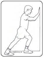
Calf muscle stretching is very useful in the treatment of many foot disorders and for the prevention of foot problems. The typical runners stretch, by leaning into a wall, is helpful. An alternative method of stretching is to stand approximately two feet from a wall. Facing the wall, turn your feet inward so you are pigeon toed. Lean forward into the wall keeping your heels on the floor and the knees extended. Also keep your back straight and do not bend at the hips. Hold the stretch for 10 seconds and do the stretch ten times in a row. Do the stretching three times each day. Always stretch the calf muscles following any form of exercise. If the stretching causes pain it should not be performed. The stretch should be felt in the body of the muscle not in the Achilles tendon or the back of the knee. If you are unable to perform the stretch properly consult with a doctor or physical trainer.

  • Apophysitis (Sever's Disease), A Cause Of Heel Pain In Children

    Calcaneal apophysitis (Sever's disease)

    Heel pain in children and adolescence: is the most common osteochondrosis (disease that affects the bone growth). Osteochondrosis is seen only in children and teens whose bones are still growing of the foot. Sever's disease or Apophysitis is a common condition that afflicts children usually between the ages of 8 to 15 years old.  Often this is confused with plantar fasciitis which is rare in children.  This is a condition of inflammation of the heel's growth plates. 

    The spontaneous development of pain in children generally indicates some form of injury to the growth plate of a growing bone. This can occur without a specific memorable event. When pain occurs in the heel of a child the most likely cause is due to injury of the growth plate in the heel bone. This is called Sever's disease. A condition that may mimic Seiver's disease is Achilles tendonitis. Achilles tendonitis is inflammation of the tendon attached to the back of the heel. A tight Achilles tendon may contribute to Sever's disease by pulling excessively on the growth plate of the heel bone. It is frequently seen in the active soccer, football or baseball player. Sport shoes with cleats seem to aggravate the condition. It is believed that the condition is due to an underlying mechanical problem with the way the foot functions.

    CalcanealApophysitis

    Underlying anatomy: This is a condition that affects the cartilage growth plate and the separate island of growing bone on the back of the heel bone. This growth plate is called the physeal plate. The island of growing bone is called the apophysis. It has the insertion attachment of the Achilles tendon, and the attachment of the plantar fascia.  This island of bone is under traction from both of these soft tissue tendon and tendon-like attachments. 

    Causes of Sever’s disease: Mechanically, the heel takes a beating. And the apophyseal bone is located near the point of impact for the heel bone at heel strike and with most weight bearing activities. This includes running, jumping and walking. Heavy impact activities like soccer, football and gymnastics are commonly associated with this problem. In addition to this, there is traction on this apophyseal bone and the associated physeal line of growth cartilage. This traction on the apopysis (island of bone) along with the impact of weight bearing activities can lead to inflammation and pain. Tight Achilles and calf muscles also can contribute to this problem, and why stretching is discussed later.

    Additional factors: Having flatfeet or very pronated feet can make one prone to Sever's disease. But also patient’s that have a very high arch foot structure tend to have a very high shock and high impact heel strike. This also puts extra stress on the heel and apophysis.

    Symptoms of Sever's disease: Symptoms include heel pain related to sports activities and worsen after those sport and exercise activities.  However, some children who are not in a sport may also get this if they are physically active. If you notice that your child is “walking on their toes” this is a sign of possible heel pain. The pain is usually on the back of the heel, the sides of the heel, the bottom of the heel, or a combination of all of these. We typically don't see swelling with this, however if pressure is applied to the sides of the heel pain may be reported. Sometimes the pain is so bad the child will have to limp, or take a break from sports activity either for a few days or few months.

    Diagnostic evaluation: This can include physical examination and x-ray evaluation. X-rays may show some increased density or sclerosis of the apophysis (island of bone on the back of the heel). This problem may be on one side or bilateral.

    Treatment of Sever’s disease:  Home treatment consists of calf muscle stretching exercises, heel cushions in the shoes, and/or oral anti-inflammatory medications like Tylenol or Advil. Icing the area may provide some temporary relief. If the condition persists the child should be evaluated by a podiatrist for abnormal foot function. In severe cases a below the knee walking cast may be required. Treatment may require the use of custom-made shoe inserts called orthotics. Orthotics work by correcting foot function and will fit into most normal shoes and athletic cleats.

    • Rest
      • Severe cases will need to be treated with a cast boot.
      • Take a break from sport activity until the pain has significant improvement.
      •  If the problem is bad enough, it is important to totally rest the symptomatic foot.
    • Anti-inflammatory treatments:
      • Icing
      • Over-the-counter anti-inflammatory medicine as recommended by your pediatrician or podiatrist.
    • Shock absorption and support
      • Don't go barefoot at home, wear some type of good sandal or shoe
      • A significant and/or chronic case should be treated with prescription orthotics          
               This addresses mechanical problems that cause this problem
      • Using an over-the-counter heel cushion inside of the shoe
      • Athletic foot taping
    • Stretching
      • Runners stretch to stretch out the calf muscle
      • A night splint will also help
    • Severe or chronic cases
      • Respond best to prescription orthotics with specific modifications for this problem
      • May require a night splint
      • Daytime braces that may also help

    Apophysitis

     The above x-ray show fragmentation and sclerosis of the heel often seen with apophysitis.

    We treat children with foot and ankle deformities and have established protocols to deal with heel pain.  Most children respond well to stretching exercises and prescription orthotics, Orthotic Rx Center.

    Is your child suffering from heel pain? We can help.

  • Capsulitis Of The Second Toe

    What is Capsulitis of the Second Toe?
    Ligaments surrounding the joint at the base of the second toe form a “capsule,” which helps the joint to function properly. Capsulitis is a condition in which these ligaments have become inflamed.

    Although capsulitis can also occur in the joints of the third or fourth toes, it most commonly affects the second toe. This inflammation causes considerable discomfort and, if left untreated, can eventually lead to a weakening of surrounding ligaments that can cause dislocation of the toe. Capsulitis—also referred to as predislocation syndrome—is a common condition that can occur at any age.

    Causes
    It is generally believed that capsulitis of the second toe is a result of abnormal foot mechanics, where the ball of the foot beneath the toe joint takes an excessive amount of weight-bearing pressure.

    Certain conditions or characteristics can make a person prone to experiencing excessive pressure on the ball of the foot. These most commonly include a severe bunion deformity, a second toe longer than the big toe, an arch that is structurally unstable, and a tight calf muscle.

    Symptoms
    Because capsulitis of the second toe is a progressive disorder and usually worsens if left untreated, early recognition and treatment are important. In the earlier stages—the best time to seek treatment—the symptoms may include: 

    • Pain, particularly on the ball of the foot. It can feel like there’s a marble in the shoe or a sock is bunched up
    • Swelling in the area of pain, including the base of the toe
    • Difficulty wearing shoes
    • Pain when walking barefoot

    In more advanced stages, the supportive ligaments weaken leading to failure of the joint to stabilize the toe. The unstable toe drifts toward the big toe and eventually crosses over and lies on top of the big toe—resulting in “crossover toe,” the end stage of capsulitis. The symptoms of crossover toe are the same as those experienced during the earlier stages. Although the crossing over of the toe usually occurs over a period of time, it can appear more quickly if caused by injury or overuse.

    Diagnosis
    An accurate diagnosis is essential because the symptoms of capsulitis can be similar to those of a condition called Morton’s neuroma, which is treated differently from capsulitis.     

    In arriving at a diagnosis, the foot and ankle surgeon will examine the foot, press on it, and maneuver it to reproduce the symptoms. The surgeon will also look for potential causes and test the stability of the joint. X-rays are usually ordered, and other imaging studies are sometimes needed.

    Non-surgical Treatment
    The best time to treat capsulitis of the second toe is during the early stages, before the toe starts to drift toward the big toe. At that time, non-surgical approaches can be used to stabilize the joint, reduce the symptoms, and address the underlying cause of the condition. 

    The foot and ankle surgeon may select one or more of the following options for early treatment of capsulitis:

    • Rest and ice. Staying off the foot and applying ice packs help reduce the swelling and pain. Apply an ice pack, placing a thin towel between the ice and the skin. Use ice for 20 minutes and then wait at least 40 minutes before icing again.
    • Oral medications. Nonsteroidal anti-inflammatory drugs (NSAIDs), such as ibuprofen, may help relieve the pain and inflammation.
    • Taping/splinting. It may be necessary to tape the toe so that it will stay in the correct position. This helps relieve the pain and prevent further drifting of the toe.  The Dr may provide you with a temporary splint. 
    • Stretching. Stretching exercises may be prescribed for patients who have tight calf muscles.  A night splint can be employed as well.
    • Shoe modifications. Supportive shoes with stiff soles are recommended because they control the motion and lessen the amount of pressure on the ball of the foot
    • Prescription Orthotic devices. Custom orthotic devices that fit into the shoe provide support for the arch, and may play a role in preventing future symptoms.  These are not over the counter inserts these are prescription medical devices made by a physician. 
    • No barefeet.  We highly recommend not going barefoot around the house, Vionic slippers or flip-flops work extremely well.
    • Cortisone Injections.  These are often very useful in alleviating symptoms
    • Immobilization. In some severe cases of pediatric heel pain, a cast may be used to promote healing while keeping the foot and ankle totally immobile.

    When is Surgery Needed?
    Once the second toe starts moving toward the big toe, it will never go back to its normal position unless surgery is performed. The foot and ankle surgeon will select the procedure or combination of procedures best suited to the individual patient.

5 out of 5 stars
Total Reviews : 232