Information for WA Patients on Common Foot and Ankle Issues | Issaquah Foot & Ankle Specialists

Information is key to treating your foot injury. In our library, we explain the common causes of many foot conditions, including bunions, broken toes, plantar fasciitis, ingrown toenails, and more. Search through our articles to find out more about your condition.
  • Freiberg's Disease

    The spontaneous development of pain in children generally indicates some form of injury to the growth plate of a growing bone. This can occur without a specific memorable event. When pain occurs in the ball of a child's foot the most likely cause is injury to the growth plate of one of the long bones behind the toes called metatarsals. The most common bone involved is the metatarsal behind the second toe. When numbering the toes the big toe is the first toe.

    This condition is called Freiberg's disease. This disorder is most frequently seen in the adolescent between the ages of 13 - 15 years of age. It is three times as likely to occur in females as compared to males. The pain is a result of a loss of blood flow to the growth plate in the bone.

    Diagnosis

    The diagnosis of Friebergs disease is made by x-ray evaluation. X-rays will reveal a flattening of the head of the metatarsal bone. Early changes may be very subtle or not apparent. On physical exam there may be swelling in the area that is tender to touch. A useful tool for diagnosing growth plate injuries and stress fracture is to strike a tuning fork and placing the vibrating tuning fork on the area of the suspected site of injury. Pain with vibration may indicate bone or growth plate injury.

    Treatment

    Treatment consists of reducing pressure under the affected bone. This may consist of anything from using crutches to a custom insole for the shoe called an orthotic. Left untreated the affected bone may not develop properly and permeate damage to the joint behind the affect toe may result in painful arthritis. Once the joint becomes arthritic surgery to place an artificial joint may be required.

  • Sesamoids
    Sesamoids are bones embedded in a tendon and are recognized as two pea-shaped bones located in the ball of the foot, beneath the big toe joint.  Injuries to the sesamoids can involve the bones, tendons and/or surrounding tissue of the joint.  They are often caused by individuals participating in physical activities that require increased pressure on the ball of the foot, such as running, basketball, football, golf, tennis and ballet.  Individuals with high arches are at risk for developing sesamoids as well as frequent high-heeled shoe wearers.  There are three types of sesamoid injuries that can occur in the foot: turf toe, fracture and sesamoiditis.  Turf toe is an injury to the soft tissue surrounding the big toe joint and usually occurs when the big toe is extended beyond its normal range.  Symptoms include immediate, sharp pain and swelling.  Sometimes a pop is felt at the moment of injury.  Fracture is a sesamoid bone can be acute or chronic.  An acute fracture is caused by trauma, produces immediate pain and swelling, but does not affect the entire big toe joint.  A chronic fracture is a stress fracture that produces longstanding pain in the ball of the foot behind the big toe joint and is aggravated with activity and relieved with rest.  Sesamoiditis is an overuse injury involving chronic inflammation of the sesamoid bones and the tendons involved with those bones.  It's caused by increased pressure to the sesamoids and often produces a dull, longstanding pain beneath the big toe joint.  Treatment of sesamoids vary, but can include: padding, strapping or taping, steroid injections, orthotics, oral medications, physical therapy or immobilization.  In some cases, surgery may be recommended.    
  • Posterior Tibial Tendonitis

    Posterior tibial tendonitis refers to inflammation of the tibial tendon, which is located in the back portion of the lower leg.  This condition can occur through a variety of physical activities that are either performed incorrectly or with excess.  These activities can include dancing, running or swimming.  This condition can also occur when there's been some type of trauma to the lower part of the body, such as a car accident.  Patients that suffer from arthritis are also susceptible to posterior tibial tendonitis.  One of the most common ways to acquire this condition is through improperly walking.  Symptoms of posterior tibial tendonitis include: pain or edema near the arch of the foot or inner side of the ankle; pain that increases in severity as the individual rises up on the ball of the foot or when the foot is extended in the upright position; or tiredness in the foot after minimal activity.  Treatment of posterior tibial tendonitis can include resting the foot, bracing or casting the foot to protect it when walking, physical therapy or in some cases, surgery.

  • Posterior Tibial Tendon Dysfunction (PTTD)
    Posterior tibial tendon dysfunction (PTTD) is a condition caused by changes in the tendon, impairing its ability to support the arch, which results in flattening of the foot.  PTTD is often referred to as "adult acquired flatfoot" because it is the most common type of flatfoot developed during adulthood.  PTTD is normally caused by overuse of the posterior tibial tendon, which is commonly found in those that participate in physical activities such as running, walking, hiking or climbing stairs.  The symptoms of PTTD can include redness, swelling, pain, flattening of the arch and an inward rolling of the ankle.  The symptoms can change as the condition progresses.  There are many treatment options available for PTTD, including orthotics, physical therapy, medications, shoe modifications and immobilization.  Occasionally, surgery is required.
  • Heel Pain: Heel Spurs
    Heel spurs are growths of bone that extends from the heel bone.  The spurs themselves have no feeling, but they extend from the heel bone into the soft tissue surrounding the bottom of the foot, causing inflammation and pain. They are commonly characterized by a sharp poking which is localized to the heel or underneath the heel.  The pain can become very severe and bruising may occur in some cases.  Pain is found to most commonly occur after prolonged periods of rest, when taking a step or when putting pressure on the heel.  In most cases, heel spurs occur in individual over the age of 40 due to sudden weight gain, improperly fitted shoewear, pronation, gout and rheumatoid arthritis.  There are many treatment options for heel spurs, such as heel pads, arch supports, orthotic therapy and anti-inflammatory medications. In some cases, surgery is recommended.
  • Flexible Flatfoot

    What Is Flatfoot?
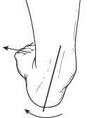
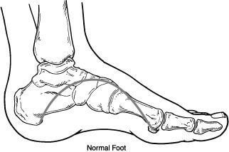
    PTTD2Flatfoot is often a complex disorder, with diverse symptoms and varying degrees of deformity and disability. There are several types of flatfoot, all of which have one characteristic in common: partial or total collapse (loss) of the arch.

    Other characteristics shared by most types of flatfoot include:

    • “Toe drift,” in which the toes and front part of the foot point outward
    • The heel tilts toward the outside and the ankle appears to turn in
    • A tight Achilles tendon, which causes the heel to lift off the ground earlier when walking and may make the problem worse
    • Bunions and hammertoes may develop as a result of a flatfoot.


    Flexible Flatfoot
    flatfoot3Flexible flatfoot is one of the most common types of flatfoot. It typically begins in childhood or adolescence and continues into adulthood. It usually occurs in both feet and progresses in severity throughout the adult years. As the deformity worsens, the soft tissues (tendons and ligaments) of the arch may stretch or tear and can become inflamed.

    The term “flexible” means that while the foot is flat when standing (weight-bearing), the arch returns when not standing.


    Symptoms

    Symptoms, which may occur in some persons with flexible flatfoot, include:

    • Pain in the heel, arch, ankle, or along the outside of the foot
    • “Rolled-in” ankle (over-pronation)
    • Pain along the shin bone (shin splint)
    • General aching or fatigue in the foot or leg
    • Low back, hip or knee pain.


    Diagnosis
    In diagnosing flatfoot, the foot and ankle surgeon examines the foot and observes how it looks when you stand and sit. X-rays are usually taken to determine the severity of the disorder. If you are diagnosed with flexible flatfoot but you don’t have any symptoms, your surgeon will explain what you might expect in the future.


    Non-surgical Treatment
    If you experience symptoms with flexible flatfoot, the surgeon may recommend non-surgical treatment options, including:

    • Activity modifications. Cut down on activities that bring you pain and avoid prolonged walking and standing to give your arches a rest.
    • Weight loss. If you are overweight, try to lose weight. Putting too much weight on your arches may aggravate your symptoms.
    • Orthotic devices. Your foot and ankle surgeon can provide you with custom orthotic devices for your shoes to give more support to the arches.
    • Immobilization. In some cases, it may be necessary to use a walking cast or to completely avoid weight-bearing.
    • Medications. Nonsteroidal anti-inflammatory drugs (NSAIDs), such as ibuprofen, help reduce pain and inflammation.
    • Physical therapy. Ultrasound therapy or other physical therapy modalities may be used to provide temporary relief.
    • Shoe modifications. Wearing shoes that support the arches is important for anyone who has flatfoot.


    When is Surgery Necessary?
    In some patients whose pain is not adequately relieved by other treatments, surgery may be considered. A variety of surgical techniques is available to correct flexible flatfoot, and one or a combination of procedures may be required to relieve the symptoms and improve foot function.

    In selecting the procedure or combination of procedures for your particular case, the foot and ankle surgeon will take into consideration the extent of your deformity based on the x-ray findings, your age, your activity level, and other factors. The length of the recovery period will vary, depending on the procedure or procedures performed.

  • Erythromelalgia
    Erythromelalgia is a fairly rare disorder manifested by vasodilatation of the blood vessels in the feet. Normally blood flow through the feet and hands is regulated by nerves and muscles in the walls of the blood vessels that either tighten and shrink the vessel's diameter so as to restrict flow or to open the blood vessels diameter to allow for more flow. This is all controlled automatically by the body and is necessary to preserve or give up body heat so that we maintain a constant body temperature.

    For instance, when we are subjected to cold temperatures the blood vessels will constrict and shunt blood back to the heart and body cavity. This is an example of how our body responds to cold and is a survival mechanism to keep us alive if we where exposed to prolonged or severely cold temperatures. The heat of the blood is not allowed to escape in the fingers and toes into the air or water around us and is sent back to the heart to keep the core body temperature warm so your heart will continue to beat. Certainly you have heard "Cold hands, warm heart". When we are hot just the opposite happens and the blood vessels enlarge or dilate allowing for more blood to go to the fingers and toes thereby 'giving up heat' to the surrounding air. The 'cooler blood then goes back to the heart where it helps lower core temperature.

    The vasoconstriction (tightening of the blood vessels) and the vasodilatation (opening of the blood vessels) is always changing and adjusting to maintain blood pressure, control body heat, regulate heart rate, among other functions you don't even think about. Part of the controls for this are partially understood and maintained by the primitive part of our nervous system called the autonomic nervous system. It is this part of our nervous system that is responsible for out heart to beat and for us to breath regularly without having to think about it.

    When these controls fail to operate normally we see the pathologic disease patterns of erythromelalgia or Raynauds disease or phenomenon.Raynauds disease or phenomenon is a vasoconstriction of the blood vessels in the feet and hands. We notice it when our fingers and toes get icy cold and turn blue or even white. This can be a very painful condition depending on how long we are subjected to the cold and the vasoconstriction since the tissues of the hands or feet are deprived of blood and therefore oxygen. In many individuals it may be very mild and not be a problem. All of us experience vasoconstriction to some degree when we are in cold weather. In the disease state however the vasoconstriction does not entirely reverse when subjected to warmer temperatures and a chronic painful situation ensues.

    Erythromelalgia on the other hand is just the opposite. The blood vessels are open or dilated and the oxygen and heat of the blood is discharged into the tissues making them turn red and feel hot all the time. Likewise this can be a very painful condition. It is a more rare disorder and less understood than Raynauds.

    Diagnosis

    Before treatment, the diagnosis should be confirmed. This can be accomplished by a variety of different medical specialties. Internal medicine or an internist is a good place to start to make sure there are no disease factors or other medications causing the Raynauds or erythromelalgia. In many instances the internist will treat the disorder so he may help you monitor medications that have undeliverable side effects or react with other medications.

    Treatment

    These disease states can be treated with varying degrees of success using drugs that induce vasodilatation or vasoconstriction. Unfortunately, the side effects of vasodilators or vasoconstrictors are often times worse than the disease. Obviously, avoiding temperatures or situations that can trigger the responses are also useful. For instance, people with vasoconstrictive problems should wear socks and well insulated shoes to maintain heat. Patients with vasodilatation problems may be more comfortable in sandals, going barefoot, or certainly using a light shoe that can 'breath' to allow heat to escape. In severe conditions pain medications can be a useful adjunct. Hypnosis and biofeedback may have some degree of success in certain individuals if administered by appropriately trained individuals.

  • Equinus

    What is Equinus?

    Equinus is a condition in which the upward bending motion of the ankle joint is limited. Someone with equinus lacks the flexibility to bring the top of the foot toward the front of the leg. Equinus can occur in one or both feet. When it involves both feet, the limitation of motion is sometimes worse in one foot than in the other.
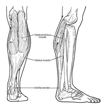
    EquinusPeople with equinus develop ways to "compensate" for their limited ankle motion, and this often leads to other foot, leg, or back problems. The most common methods of compensation are flattening of the arch or picking up the heel early when walking, placing increased pressure on the ball of the foot. Other patients compensate by "toe walking," while a smaller number take steps by bending abnormally at the hip or knee.


    Causes
    There are several possible causes for the limited range of ankle motion. Often it is due to tightness in the Achilles tendon or calf muscles (the soleus muscle and/or gastrocnemius muscle). In some patients, this tightness is congenital (present at birth) and sometimes it is an inherited trait. Other patients acquire the tightness from being in a cast, being on crutches, or frequently wearing high-heeled shoes. In addition, diabetes can affect the fibers of the Achilles tendon and cause tightness.

    Sometimes equinus is related to a bone blocking the ankle motion. For example, a fragment of a broken bone following an ankle injury, or bone block, can get in the way and restrict motion.

    Equinus may also result from one leg being shorter than the other.

    Less often, equinus is caused by spasms in the calf muscle. These spasms may be signs of an underlying neurologic disorder.


    Foot Problems Related to Equinus
    Depending on how a patient compensates for the inability to bend properly at the ankle, a variety of foot conditions can develop, including:

    • Plantar fasciitis (arch/heel pain)
    • Calf cramping
    • Tendonitis (inflammation in the Achilles tendon)
    • Metatarsalgia (pain and/or callusing on the ball of the foot)
    • Flatfoot
    • Arthritis of the midfoot (middle area of the foot)
    • Pressure sores on the ball of the foot or the arch
    • Bunions and hammertoes
    • Ankle pain
    • Shin splints


    Diagnosis
    Most patients with equinus are unaware they have this condition when they first visit the doctor. Instead, they come to the doctor seeking relief for foot problems associated with equinus.

    To diagnose equinus, the foot and ankle surgeon will evaluate the ankle's range of motion when the knee is flexed (bent) as well as extended (straightened). This enables the surgeon to identify whether the tendon or muscle is tight and to assess whether bone is interfering with ankle motion. X-rays may also be ordered. In some cases, the foot and ankle surgeon may refer the patient for neurologic evaluation.


    Non-Surgical Treatment
    Treatment includes strategies aimed at relieving the symptoms and conditions associated with equinus. In addition, the patient is treated for the equinus itself through one or more of the following options:

    • Night splint. The foot may be placed in a splint at night to keep it in a position that helps reduce tightness of the calf muscle.
    • Heel lifts. Placing heel lifts inside the shoes or wearing shoes with a moderate heel takes stress off the Achilles tendon when walking and may reduce symptoms.
    • Arch supports or prescription custom orthotics devices. Prescription custom orthotics devices that fit into the shoe are often prescribed to keep weight distributed properly and to help control muscle/tendon imbalance.
    • Physical therapy. To help remedy muscle tightness, exercises that stretch the calf muscle(s) are recommended.


    When is Surgery Needed?
    In some cases, surgery may be needed to correct the cause of equinus if it is related to a tight tendon or a bone blocking the ankle motion. The foot and ankle surgeon will determine the type of procedure that is best suited to the individual patient.

  • Enchondroma

    A subungual exostosis is a bony prominence that can occur under the toenail. They generally are a result of some form of trauma to the toe that results in the formation of bony irregularity or prominence. When they are symptomatic, removal of the spur is the treatment of choice. Additionally other small tumors called osteochondromas and enchondroma can also form in the bone beneath the toenail as well as in other bones in the body.

    An osteochondroma is a benign bone tumor that accounts for 50% of all benign bone tumors. They have a predilection for the long tubular bones of adolescents and young adults. Its peak incidence is in the second decade of life with a male to female ratio of approximately 2:1. They are generally painless or minimally painful unless they cause irritation to the surrounding tissue. When they are in the bone beneath the toenail they can deform the toenail and cause an ingrown toenail. The treatment of symptomatic osteochondromas is surgical excision. The final diagnosis is made after the bone tumor is removed and examined by a pathologist. Recurrence of the tumor is possible following their removal. Hereditary multiple exostosis (osteochondromatosis) have a prominent hereditary incidence which affects males more often then females. The disease is characterized by the presence of multiple exostosis, which are frequently bilateral and somewhat symmetrical and usually make their appearance during childhood or adolescence.

    Enchondroma is a fairly common benign cartilaginous tumor, which is the most common bone tumor of the hands and feet. They affect patients in a wide age range with no sex predilection. When they occur within the small tubular bones the tumor can involve large portions of these bones, causing thinning of the cortex of the bone. This can weaken the bone and cause it to break spontaneously. When they occur in the small bone in the end of the toe they can cause pain that may mimic the pain of ingrown toenails. Ollier's disease, also known as enchondromatosis, shows a strong predilection for the small bones in the hands and toes (phalanges) and the long bones behind the phalanges called metatarsals. It is often shows an asymmetric involvement, tending to affect one side of the body more than the other and has a propensity to transform into a malignant sarcoma. Maffucci's syndrome is a very rare form of enchondromatosis associated with multiple soft tissue hemangiomas.This tumor has a greater predilection for the hands and feet, and has a greater tendency toward malignant transformation than Ollier's disease.

  • Electromyography (EMG) / Nerve Conduction Velocity (NCV)

    This test is used to test the nerves and muscles in your entire lower extremity. Your doctor will usually order this test when he suspects that there may be some type of problem with the nerve supply to your foot and leg. Commonly the EMG/NCV test is used to diagnosis one of the following: Tarsal Tunnel Syndrome, Peripheral Neuropathy, Neuromuscular disorders, Nerve palsy or Paralysis, and Radioculopathy. Your doctor typically will refer you to either a hospital or a neurologist to have the test performed.

    The EMG portion of the test is used to record the electrical activity in your muscles. It can diagnose diseases of the nerves and muscles. It can detect conditions such as tarsal tunnel syndrome, inflamed muscles and pinched nerves. A tiny needle, called an electrode, is inserted directly into a specific muscle belly. The electrode then records the activity during the insertion, while the muscle is at rest, and while the muscle contracts. Nerve and muscle diseases alter the pattern of electrical activity in these muscles, which is record both audibly and on a computer screen. After the first muscle is tested, the electrode may be inserted into another muscle. Muscles chosen for the testing vary with the patient's symptoms and may be modified, depending on the results from the first muscles tested. Total testing time may range from just a few minutes to more than an hour, depending upon how many muscles are tested. After the exam, you may feel tenderness in the tested muscles. There is a slight risk of minor, localized inflammation in muscles during the test. This usually lasts only a few hours. Other common patient complaints are pain with insertion of the electrode.

    Most of the time the Nerve Conduction Velocity Test will accompany the EMG Test. The NCV evaluates the health of the peripheral nerve by recording how fast an electrical impulse travels through it. A peripheral nerve transmits information between the spinal cord and the muscles. You will be resting on a cart or bed and electrodes will be taped to your skin. A stimulator will be held against your skin, which sends out a small electrical charge along the nerve. You may feel a tingle or your muscles may twitch but this shock is not harmful. Each test will take only a few minutes. After the exam the electrodes will be removed and your skin cleaned. The time between the stimulation and response will be recorded to determine how quickly and thoroughly that the impulse is sent. A number of nervous system diseases may reduce the speed of this impulse. Each nerve test takes just a few minutes to an hour, depending upon how many nerves are being tested.

    While the hospital or neurologist's office will give you instructions for the day of the examination, a few general preparations will help. Eat normally and take medication as you usually would. If you are taking a blood thinner, make sure you inform the testing facility and ask the ordering physician about the use of the medication and the timing of the test. Bath or shower the morning of the examination. Avoid bath oils or any skin lotions or emollients the day of the examination.

    A typical EMG/NCV of the lower extremity takes approximately 45 minutes. This test is an important tool for diagnosing diseases of the nervous system, you can help ensure the best results if you relax and cooperate with the technicians. Make sure that you ask any questions that you have about the test before it is performed. Your physician will discuss the results with you. If you have any further questions regarding why this test was ordered for you, please ask your physician.

5 out of 5 stars
Total Reviews : 232