Heel Pain

  • Calf Muscle Stretching

    Excessive tightness of the calf muscles can contribute to many foot problems and some knee problems. The Achilles tendon attaches the calf muscle to the back of the heel. As the calf muscle tightens up it limits the movement of the ankle joint. When there is not adequate range of motion at the ankle during walking or exercising the foot joints are forced to move in an abnormal fashion. The foot is forced to flatten and the forces generated into the ball of the foot are extreme in nature. Over time, this repeated strain results in a variety of foot problems. The calf muscle has a natural tendency to tighten, as we get older. Exercise also causes the muscle to tighten.

    calf stretchCalf muscle stretching is very useful in the treatment of many foot disorders and for the prevention of foot problems. The typical runners stretch, by leaning into a wall, is helpful. An alternative method of stretching is to stand approximately two feet from a wall. Facing the wall, turn your feet inward so you are pigeon toed. Lean forward into the wall keeping your heels on the floor and the knees extended. Also keep your back straight and do not bend at the hips. Hold the stretch for 10 seconds and do the stretch ten times in a row. Do the stretching three times each day. Always stretch the calf muscles following any form of exercise. If the stretching causes pain it should not be performed. The stretch should be felt in the body of the muscle not in the Achilles tendon or the back of the knee. If you are unable to perform the stretch properly consult with a doctor or physical trainer.

  • Capsulitis Of The Second Toe

    What is Capsulitis of the Second Toe?
    Ligaments surrounding the joint at the base of the second toe form a “capsule,” which helps the joint to function properly. Capsulitis is a condition in which these ligaments have become inflamed.

    Although capsulitis can also occur in the joints of the third or fourth toes, it most commonly affects the second toe. This inflammation causes considerable discomfort and, if left untreated, can eventually lead to a weakening of surrounding ligaments that can cause dislocation of the toe. Capsulitis—also referred to as predislocation syndrome—is a common condition that can occur at any age.

    Causes
    It is generally believed that capsulitis of the second toe is a result of abnormal foot mechanics, where the ball of the foot beneath the toe joint takes an excessive amount of weight-bearing pressure.

    Certain conditions or characteristics can make a person prone to experiencing excessive pressure on the ball of the foot. These most commonly include a severe bunion deformity, a second toe longer than the big toe, an arch that is structurally unstable, and a tight calf muscle.

    Symptoms
    Because capsulitis of the second toe is a progressive disorder and usually worsens if left untreated, early recognition and treatment are important. In the earlier stages—the best time to seek treatment—the symptoms may include: 

    • Pain, particularly on the ball of the foot. It can feel like there’s a marble in the shoe or a sock is bunched up
    • Swelling in the area of pain, including the base of the toe
    • Difficulty wearing shoes
    • Pain when walking barefoot

    In more advanced stages, the supportive ligaments weaken leading to failure of the joint to stabilize the toe. The unstable toe drifts toward the big toe and eventually crosses over and lies on top of the big toe—resulting in “crossover toe,” the end stage of capsulitis. The symptoms of crossover toe are the same as those experienced during the earlier stages. Although the crossing over of the toe usually occurs over a period of time, it can appear more quickly if caused by injury or overuse.

    Diagnosis
    An accurate diagnosis is essential because the symptoms of capsulitis can be similar to those of a condition called Morton’s neuroma, which is treated differently from capsulitis.     

    In arriving at a diagnosis, the foot and ankle surgeon will examine the foot, press on it, and maneuver it to reproduce the symptoms. The surgeon will also look for potential causes and test the stability of the joint. X-rays are usually ordered, and other imaging studies are sometimes needed.

    Non-surgical Treatment
    The best time to treat capsulitis of the second toe is during the early stages, before the toe starts to drift toward the big toe. At that time, non-surgical approaches can be used to stabilize the joint, reduce the symptoms, and address the underlying cause of the condition. 

    The foot and ankle surgeon may select one or more of the following options for early treatment of capsulitis:

    • Rest and ice. Staying off the foot and applying ice packs help reduce the swelling and pain. Apply an ice pack, placing a thin towel between the ice and the skin. Use ice for 20 minutes and then wait at least 40 minutes before icing again.
    • Oral medications. Nonsteroidal anti-inflammatory drugs (NSAIDs), such as ibuprofen, may help relieve the pain and inflammation.
    • Taping/splinting. It may be necessary to tape the toe so that it will stay in the correct position. This helps relieve the pain and prevent further drifting of the toe.  The Dr may provide you with a temporary splint. 
    • Stretching. Stretching exercises may be prescribed for patients who have tight calf muscles.  A night splint can be employed as well.
    • Shoe modifications. Supportive shoes with stiff soles are recommended because they control the motion and lessen the amount of pressure on the ball of the foot
    • Prescription Orthotic devices. Custom orthotic devices that fit into the shoe provide support for the arch, and may play a role in preventing future symptoms.  These are not over the counter inserts these are prescription medical devices made by a physician. 
    • No barefeet.  We highly recommend not going barefoot around the house, Vionic slippers or flip-flops work extremely well.
    • Cortisone Injections.  These are often very useful in alleviating symptoms
    • Immobilization. In some severe cases of pediatric heel pain, a cast may be used to promote healing while keeping the foot and ankle totally immobile.

    When is Surgery Needed?
    Once the second toe starts moving toward the big toe, it will never go back to its normal position unless surgery is performed. The foot and ankle surgeon will select the procedure or combination of procedures best suited to the individual patient.

  • New Treatment: Dry Needling

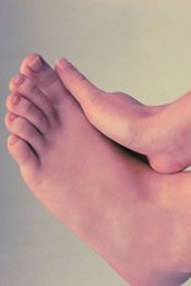
    New treatment for plantar fasciitis: Dry needling combined with a cortisone injection, guided ultrasound imaging-the alternative to plantar fasciitis surgery. Researchers state that this approach has a 95% success rate. They also state that the ultrasound guidance makes the procedure very safe. They recommend the procedure combined with orthotic treatment and advised that 42/44 of their study patients were cured of their symptoms within 2-3 weeks of treatment. The dry needling technique allows for increased blood flow to the symptomatic plantar fascia region. This helps it heal faster. Traditionally structures like the plantar fascia, tendons and ligaments do not have a lot of blood flow, so they tend to heal slowly. The premise behind this is quite similar to the APC percutaneous growth factor procedure that we also utilize. One difference is that this new procedure is much less costly and can be done right in our exam rooms with local anesthetic.

    Our doctors have been safely performing ultrasound-guided cortisone injections for plantar fasciitis for several years. This approach has been successful when combined with other types of treatment such as prescription orthotics. We now offer this new technique at our clinic with the dry needling technique combined with a cortisone injection and we further recommend that the foot be in a protective medical boot for 3 weeks following the procedure.

  • Dry Needling And Plantar Fasciitis: Part Two

    What to expect
    At Issaquah Foot and Ankle Specialists, we have developed a treatment protocol and have fine tuned the technique for dry needling for plantar fasciitis. In addition, we have developed adjunctive treatments that help further ensure that chronic plantar fasciitis is resolved. Dr. Young has personally communicated with Dr. Lucia Sconfienza by e-mail soon after the results were first presented. He discussed details of the procedure to make certain that we were able to provide our patients with the best possible treatment and outcome.


    Who is a candidate for dry needling
    Those individuals who have already tried other means of treatment for plantar fasciitis including but not limited to prescription orthotics, excellent shoes, a night splint, cortisone injections, taping and bracing.

    Diagnostic ultrasound imaging typically will show enlargement of the plantar fascia in those individuals with severe plantar fasciitis. The typical thickness of the plantar fascia at the heel is anywhere from 3 to 4 mm thick maximum.  Those individuals with chronic plantar fasciitis typically will be anywhere from 20% to even 100% thicker than normal. This means that the fascia can be 5 mm to 8 mm thick (even up to 10 mm in some severe cases). If an individual does not have significant swelling (edema) of the plantar fascia, then the prognosis for improvement with dry needling and many of the other traditional plantar fasciitis treatments tend to be less effective. In general, it is a severe case without swelling of the fascia and there are other significant causes of the heel pain instead of plantar fasciitis. 


    Dry needling description

    First, we utilize our electrical stimulation in conjunction with the local anesthetic to help minimize discomfort.  After the local anesthetic has fully taken effect, the procedure is done with no discomfort whatsoever.

    High-resolution ultrasound guidance is used to direct the procedure, which allows for the precise finding of the exact location. A needle and syringe is used to repeatedly probe the fascia throughout the thickest portion at the calcaneal attachment site.  This probing can go all the way down to the attachment and periosteum (outer layer bone). Once this has been done for the prescribed time frame, then we typically utilize a combination of Kenalog and dexamethasone phosphate at the interface between the plantar fascia in the plantar fat pad. This is another example of where the ultrasound guidance is essential. Using this technique, we can make certain that the cortisone is not injected into the plantar fascia itself or the plantar fat pad. Injecting into the plantar fascia would raise the risk of a partial tear or rupture of the plantar fascia. In addition, injecting into the plantar fat pad can cause long-term thinning (atrophy) of the fat pad.


    New treatment variations on this technique 
    This is where instead of using cortisone, we use autogenous platelet rich plasma with growth factor. It is also possible to do this procedure without using any cortisone.


    Frequently ask questions
    Is it safe?
    Yes, dry needling is very safe.  There are few risks associated with the procedure and these risks are minimal compared to surgical risks.  A patient may experience an allergic reaction from the anesthesia and they do run a small risk of post-injection infection.  Some patients may have pain for a few days following the procedure as well. 

    Is there any pain with the procedure?
    No, a patient shouldn't experience any pain during the procedure.  We use a local anesthetic to numb the foot as well as electrical stimulation with our TENS unit to make sure the procedure is pain-free. 

    How long have you been doing dry needling? 
    Our clinic started providing the service in December of 2008. Our clinic has been providing high-resolution ultrasound guided injections for over 5 years.

    Does my insurance cover this procedure? 
    We accept many different insurance plans at our office.  This is something that our billing staff can help you with.


    Post-procedure treatment protocol essentials:

    • Avoid anti-inflammatory medication and icing
    • Use a cast boot.  This is done to protect the soft tissue in the heel as it now starts the healing process.
    • Allow for bruising and inflammation.  When you tear or sprain a tendon or ligaments, there is deep internal swelling or bruising that occurs due to the need to protect the tissue which allows for soft tissue healing phases to occur. The same should occur with this procedure.


    What else can be done to help make sure this is effective:

    • Use a night splint every night for the first 2-3 months.
    • Prescription orthotics that are specific to your problem and to plantar fasciitis. You want to treat the mechanics that caused the problem to begin with.
    • Electrical stimulation may be beneficial to the post-procedure healing time frame.


    Contact us today for an appointment or to learn more.

  • Plantar Fasciitis And Exercise

    The most important thing to remember when exercising with plantar fasciitis is to use pain as your guideline. Runners can be prone to plantar fasciitis due to a developing overuse injury. Once the plantar fasciitis starts, with continued exercise or impact activity, it continues to get worse. By the time it is bad enough that something needs to be done, there is already significant underlying enlargement and swelling of the plantar fascia.

    It is important to remember that plantar fasciitis has both an inflammatory component and mechanical component.  From a mechanical standpoint, it is important to immediately stop doing impact exercise or activities. Here are some tips for relieving pain causing by plantar fasciitis:

    • No impact exercise. Low-impact activities such as aqua jogging and stationary cycling are great alternatives. 
    • Using of a night splint to help stretch out the the calf muscle.
    • Aggressive stretching, but making certain to stretch the calf, not the foot.
    • Taping the foot especially for any limited impact activities or weightbearing activities.
    • Using a Bledsoe plantar fasciitis brace
    • Replacing worn out shoes and wearing shoes with excellent support.
    • For significant cases, the use of prescription orthotics is ideal.
    • Massage therapy and ASTYM physical therapy massage

    From an anti-inflammatory standpoint, the plantar fascia itself is almost always inflamed and this inflammation can be addressed by various approaches:

    • Icing the foot for least 20 minutes directly after physical activity and in the evening
    • An over-the-counter anti-inflammatory can be utilized per label instructions. But, it is important to realize that this can mask the pain and it is possible to then overdo it because one might think that it is healed when it is not.
    • Some forms of physical therapy such as ultrasound therapy or electrical stimulationprovide an anti-inflammatory effect ORIF physically decrease deep swelling (edema).
    • cortisone injection can be beneficial. Ideally, this should be directed at the interface between the plantar fascia and the fat pad and not within the plantar fascia. An injection within the fascia increases the risk of a tear of the fascia.

    More aggressive treatments:

    Exercise guidelines:

    • Use pain as a reference to your allowable activity level.
    • When you are in the acute phase of plantar fasciitis with pronounced pain in the morning and when you get up from sitting, it still too early to return to exercise.
    • Initially, non-weightbearing exercises may be tolerated without an increase in pain that day or the following morning.
    • Use of a recumbent bike, swimming or aqua jogging

    After several weeks (or even a month or longer), the following are good exercise options: elliptical machine, rowing machine, outdoor cycling and spinning class.

    The last phase would be returning to the most pronounced impact activities such as running, basketball or other impact sports. There should be 90-95% improvement with minimal pain before returning to these impact activities.

    Relapse: 
    If you start to increase your activity level, but start having increased pain levels, you have gone too far too fast. It is time to back off very quickly. Remember that some forms of mechanical support will allow you to do more and pay less of a price.

  • Adult Flatfoot

    Flatfoot is a condition in which the longitudinal arch in the foot, which runs lengthwise along the sole of the foot, has not developed correctly and is lowered or flattened out.  There are many causes of flatfoot, such as: genetics, rheumatoid arthritis, stroke or diabetes.  Individuals who have flat feet rarely have symptoms, but they might experience pain in the following instances: sudden weight gain, minor injury, incorrectly fitted footwear, changes in work environment or excessive standing, walking, jumping or running.  Treatment for flat feet usually consists of wearing spacious, comfortable shoes with good arch support.  Many times, orthotics are recommended to balance the foot in a neutral position and cushion the foot from excessive pounding.

5 out of 5 stars
Total Reviews : 232