Children's Issues

  • Kids Feet

    Our Issaquah clinic is kid friendly! We see a large number of children from the Seattle area and beyond. There are a variety of problems that can affect their feet.

    Arch Problems and Walking Problems

    Many children start life off with low arches. We all start out life with quite a bit of "baby fat." It's surprising, but this is even in the foot and especially the instep. This makes it difficult to tell if the young child has a normal arch or not. The problem is, that sometimes they don't have a normal arch. Even in these cases, sometimes the parents are told that they will outgrow this low arch or flatfoot. However, many children do not outgrow these problems and they become lifelong challenges. We evaluate and treat a number of these different arch and walking concerns.
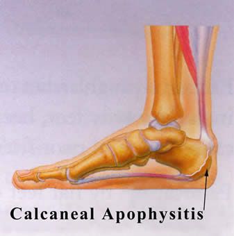
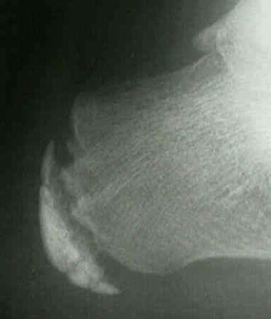
    Heel Pain

    Many of our pediatric patients have pain in their heels. This makes it difficult to do the sports they like. We have developed treatments that can eliminate this heel pain rapidly. A common cause of this heel pain is irritation to the growth plate in the heel bone (calcaneal apophysitisor Sever's Disease). Other possible causes include Achilles tendonitis and plantar fasciitis.

    Growing Pains

    Children may complain of pain in their legs during or after activity or sports. Often, this is not really growing pains but rather leg muscle pain and fatigue. These muscles support and control foot function. In adults these can be part of the shinsplint syndrome. Children can get a variation of this. Our clinic has very effective treatments for this condition.

    Children's Sports Injuries

    We also see and treat foot and ankle injuries. So many kids are doing multiple sports. This is great exercise, but it does put an extra work load on the legs and feet. This can cause problems and injuries ranging from a simple sprain or tendonitis to a fracture that is either a typical fracture or a growth plate type of injury to the bone. Each sport seems to require slightly different treatment considerations. Some of these different sports include soccer, football, basketball, lacrosse, baseball, gymnastics, dance and ballet, track and field, martial arts and volleyball.

    Plantar Warts

    These are warts on the bottom of the feet. Compared to adults, children are often barefoot, which makes them more vulnerable to these plantar warts. The virus that causes these warts only live in human skin and they can be contagious. These warts can be painful and often are much deeper than expected. What you see on the skin is often just "the tip of the iceberg." Many medical clinics treat all warts with a liquid nitrogen freezing technique; this is often very painful and causes many children to be apprehensive about treatment for plantar warts. However, we have found a number of treatments that are very effective and often do not have any associated pain. Our office has become a treatment center for resistant plantar warts. We work with many of the local pediatricians and family practice clinics to treat these problems.

    Ingrown Toenails

    Adolescent boys in particular get ingrown toenails. They are growing so fast that their shoes can become snug fitting before they get replaced. This shoe pressure is often a causative factor in ingrown toenails.

    For children, we use a combination of topical anesthetic -- ethyl chloride (cold spray), and gentle electrical stimulation. The patient controls electrical stimulation themselves to help keep the nerves "busy." Then, the local anesthetic injection is administered. Our younger patients benefit from this special technique. They are often very apprehensive about an injection, but with our techniques, the patient and their parents are usually pleasantly surprised at how well they do.

  • Freiberg's Disease

    The spontaneous development of pain in children generally indicates some form of injury to the growth plate of a growing bone. This can occur without a specific memorable event. When pain occurs in the ball of a child's foot the most likely cause is injury to the growth plate of one of the long bones behind the toes called metatarsals. The most common bone involved is the metatarsal behind the second toe. When numbering the toes the big toe is the first toe.

    This condition is called Freiberg's disease. This disorder is most frequently seen in the adolescent between the ages of 13 - 15 years of age. It is three times as likely to occur in females as compared to males. The pain is a result of a loss of blood flow to the growth plate in the bone.

    Diagnosis

    The diagnosis of Friebergs disease is made by x-ray evaluation. X-rays will reveal a flattening of the head of the metatarsal bone. Early changes may be very subtle or not apparent. On physical exam there may be swelling in the area that is tender to touch. A useful tool for diagnosing growth plate injuries and stress fracture is to strike a tuning fork and placing the vibrating tuning fork on the area of the suspected site of injury. Pain with vibration may indicate bone or growth plate injury.

    Treatment

    Treatment consists of reducing pressure under the affected bone. This may consist of anything from using crutches to a custom insole for the shoe called an orthotic. Left untreated the affected bone may not develop properly and permeate damage to the joint behind the affect toe may result in painful arthritis. Once the joint becomes arthritic surgery to place an artificial joint may be required.

  • Curled Toes

    Deformities of the toes are common in the pediatric population. Generally they are congenital in nature with both or one of the parents having the same or similar condition. Many of these deformities are present at birth and can become worse with time. Rarely do children outgrow these deformities although rare instances of spontaneous resolution of some deformities have been reported.

    Malformation of the toes in infancy and early childhood are rarely symptomatic. The complaints of parents are more cosmetic in nature. However, as the child matures these deformities progress from a flexible deformity to a rigid deformity and become progressively symptomatic. Many of these deformities are unresponsive to conservative treatment. Common digital deformities are underlapping toesoverlapping toes,flexed or contracted toes and mallet toes. Quite often a prolonged course of digital splitting and exercises may be recommended but generally with minimal gain. As the deformity becomes more rigid surgery will most likely be required if correction of the deformity is the goal.

    Underlapping Toes

    Description

    Underlapping toes are commonly seen in the adult and pediatric population. The toes most often involved are the fourth and fifth toes. A special form of underlaping toes is calledclinodactyly or congenital curly toes. Clinodactyly is fairly common and follows a familial pattern. One or more toes may be involved with toes three, four, and five of both feet being most commonly affected.

    The exact cause of the deformity is unclear. A possible etiology is an imbalance in muscle strength of the small muscles of the foot. This is aggravated by a subtle abnormality in the orientation on the joints in the foot just below the ankle joint called the subtalar joint. This results in an abnormal pull of the ligaments in the toes causing them to curl. With weight bearing the deformity is increased and a folding or curling of the toes results in the formation of callus on the outside margin of the end of the toe. Tight fitting shoes can aggravate the condition.

    Treatment

    The age of the patient, degree of the deformity and symptoms determine treatment. If symptoms are minimal, a wait and see approach is often the best bet. When treatment is indicated the degree of deformity determines the level of correction. When the deformity is flexible in nature a simple release of the tendon in the bottom of the toe will allow for straightening of the toe. If the deformity is rigid in nature then removal of a small portion of the bone in the toe may be necessary. Both of these procedures are common in the adult patient for the correction of hammertoe deformity. If skin contracture is present a derotational skin plasy may be required.

    Overlapping Toes - Overlapping Fifth Toe

    Description

    This deformity is characterized by one toe lying on top of an adjacent toe. The most common toe involved is the fifth toe. When one of the central toes is involved the second toe is most commonly affected. The etiology of the condition is not well understood. It is though that it may be caused by the position of the fetus in the womb during development. The condition my run in families so there may be a hereditary component to the deformity.

    Treatment

    Effective conservative treatment depends upon how early the diagnosis is made. In infancy, passive stretching and adhesive tapping is most commonly used. This may require 6 to 12 weeks to accomplish and reoccurrence is not uncommon. Rarely will the deformity correct itself. As the individual matures the deformity becomes fixed. When surgical correction is warranted a skin plasty is required to release the contracture of the skin associated with the deformity. Additionally a tendon release and a release of the soft tissues about the joint at the base of the fifth toe may be required. In severe cases the toe may require the placement of a pin to hold the toe in a straightened position. The pin, which exits the tip of the toe, may be left in place for up to three weeks. During this period of time the patient must curtail their activities significantly and wear either a post-operative type shoe or a removable cast. Excessive movement at the surgical site can result in a less than desirable result. The pin can be easily removed in the doctor's office with minimal discomfort. Following removal of the pin splinting of the toe may be required for an additional two to three weeks.

    Hammertoes and Mallet Toes

    Description

    Another common digital deformity is contracture of the toes in the formation of hammertoes and mallet toes. Hammertoes are described in depth in another article. Mallet toes are a result of contracture of the last joint in the toe. In the pediatric population it is often flexible and not painful. Over time the deformity becomes rigid and a callus may form on the skin overlying the joint at the end of the toe. Additionally the toenail may become thickened and deformed form the repetitive jamming of the toe while walking. The deformity usually involves one or two toes, with the second toe most commonly affected. Mallet toes have several etiologies. Longer toes that are forced against a short toe box in the shoe will, over time, develop a contracture of the last joint in the toe causing a mallet toe.

    Treatment

    Conservative treatment consists of padding and strapping the toes into a corrected position. This treatment may alleviate the symptoms but will not correct the deformity. Diabetic patients often develop ulcerations on the ends of their toes secondary to mallet toe deformity and the pressure that results from the toe jamming into the shoe. When standing, the toe will demonstrate a contracture, with the tip of the toe facing downward into the floor. If the deformity is flexible a simple release of the tendon in the bottom of the toe will allow straightening of the toe. Following the procedure the patient must avoid shoes that cause jamming of the toe or the deformity can reoccur. When the deformity is rigid surgical correction requires the removal of a small section of bone in the last joint of the toe. On occasion fusion of the last two bones in the toe may be necessary. This requires removing the cartilage from the last joint in the toe and pinning the bones together. When the bone heals it forms a single bone and the toe remains in a straightened position. Healing time is dependent upon the procedure selected. If a tendon release is performed the patient my return to a roomy shoe within a week. If the toe is straightened by removing a section of the bone in the toe it make ten days to three weeks for a patient to return to normal shoes. If a fusion is performed to straighten the toe, the patient may not return to normal shoes for 6 to 8 weeks. Time off from work will depend upon the type of shoe gear that must be worn and the level of activity necessary to perform the job. A minimum of three to four days off from work is generally recommended and longer if the job responsibilities can not be modified to accommodate the normal healing time for the surgery.

  • Apophysitis (Sever's Disease), A Cause Of Heel Pain In Children

    Calcaneal apophysitis (Sever's disease)

    Heel pain in children and adolescence: is the most common osteochondrosis (disease that affects the bone growth). Osteochondrosis is seen only in children and teens whose bones are still growing of the foot. Sever's disease or Apophysitis is a common condition that afflicts children usually between the ages of 8 to 15 years old.  Often this is confused with plantar fasciitis which is rare in children.  This is a condition of inflammation of the heel's growth plates. 

    The spontaneous development of pain in children generally indicates some form of injury to the growth plate of a growing bone. This can occur without a specific memorable event. When pain occurs in the heel of a child the most likely cause is due to injury of the growth plate in the heel bone. This is called Sever's disease. A condition that may mimic Seiver's disease is Achilles tendonitis. Achilles tendonitis is inflammation of the tendon attached to the back of the heel. A tight Achilles tendon may contribute to Sever's disease by pulling excessively on the growth plate of the heel bone. It is frequently seen in the active soccer, football or baseball player. Sport shoes with cleats seem to aggravate the condition. It is believed that the condition is due to an underlying mechanical problem with the way the foot functions.

    CalcanealApophysitis

    Underlying anatomy: This is a condition that affects the cartilage growth plate and the separate island of growing bone on the back of the heel bone. This growth plate is called the physeal plate. The island of growing bone is called the apophysis. It has the insertion attachment of the Achilles tendon, and the attachment of the plantar fascia.  This island of bone is under traction from both of these soft tissue tendon and tendon-like attachments. 

    Causes of Sever’s disease: Mechanically, the heel takes a beating. And the apophyseal bone is located near the point of impact for the heel bone at heel strike and with most weight bearing activities. This includes running, jumping and walking. Heavy impact activities like soccer, football and gymnastics are commonly associated with this problem. In addition to this, there is traction on this apophyseal bone and the associated physeal line of growth cartilage. This traction on the apopysis (island of bone) along with the impact of weight bearing activities can lead to inflammation and pain. Tight Achilles and calf muscles also can contribute to this problem, and why stretching is discussed later.

    Additional factors: Having flatfeet or very pronated feet can make one prone to Sever's disease. But also patient’s that have a very high arch foot structure tend to have a very high shock and high impact heel strike. This also puts extra stress on the heel and apophysis.

    Symptoms of Sever's disease: Symptoms include heel pain related to sports activities and worsen after those sport and exercise activities.  However, some children who are not in a sport may also get this if they are physically active. If you notice that your child is “walking on their toes” this is a sign of possible heel pain. The pain is usually on the back of the heel, the sides of the heel, the bottom of the heel, or a combination of all of these. We typically don't see swelling with this, however if pressure is applied to the sides of the heel pain may be reported. Sometimes the pain is so bad the child will have to limp, or take a break from sports activity either for a few days or few months.

    Diagnostic evaluation: This can include physical examination and x-ray evaluation. X-rays may show some increased density or sclerosis of the apophysis (island of bone on the back of the heel). This problem may be on one side or bilateral.

    Treatment of Sever’s disease:  Home treatment consists of calf muscle stretching exercises, heel cushions in the shoes, and/or oral anti-inflammatory medications like Tylenol or Advil. Icing the area may provide some temporary relief. If the condition persists the child should be evaluated by a podiatrist for abnormal foot function. In severe cases a below the knee walking cast may be required. Treatment may require the use of custom-made shoe inserts called orthotics. Orthotics work by correcting foot function and will fit into most normal shoes and athletic cleats.

    • Rest
      • Severe cases will need to be treated with a cast boot.
      • Take a break from sport activity until the pain has significant improvement.
      •  If the problem is bad enough, it is important to totally rest the symptomatic foot.
    • Anti-inflammatory treatments:
      • Icing
      • Over-the-counter anti-inflammatory medicine as recommended by your pediatrician or podiatrist.
    • Shock absorption and support
      • Don't go barefoot at home, wear some type of good sandal or shoe
      • A significant and/or chronic case should be treated with prescription orthotics          
               This addresses mechanical problems that cause this problem
      • Using an over-the-counter heel cushion inside of the shoe
      • Athletic foot taping
    • Stretching
      • Runners stretch to stretch out the calf muscle
      • A night splint will also help
    • Severe or chronic cases
      • Respond best to prescription orthotics with specific modifications for this problem
      • May require a night splint
      • Daytime braces that may also help

    Apophysitis

     The above x-ray show fragmentation and sclerosis of the heel often seen with apophysitis.

    We treat children with foot and ankle deformities and have established protocols to deal with heel pain.  Most children respond well to stretching exercises and prescription orthotics, Orthotic Rx Center.

    Is your child suffering from heel pain? We can help.

5 out of 5 stars
Total Reviews : 233