Ankle Issues

  • MRI - Magnetic Resonance Imaging

    In some foot and ankle conditions, your doctor may order an MRI to help diagnosis the problem that you are having with your foot and ankle. In the foot and ankle, MRI can be used to diagnosis the following conditions:

    1. Tendon injuries
    2. Ligament injuries
    3. Cartilage injuries
    4. Fractures
    5. Tumors (soft tissue and bone)
    6. Infection
    7. Avascular necrosis
    8. Non-unions or delayed unions of bone fractures
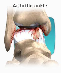
    9. Arthritis

    What is MRI?

    MRI is the newest way to view the human body since the CT (CAT) scanner was invented. Unlike CT scans, MRI does not use radiation in the conventional sense of the word. Rather, it combines the use of a large magnet and radio waves. The hydrogen atoms in the patient's body react to the magnetic field, and a computer analyzes the results and makes pictures of the inside of your body. MRI pictures show the soft tissues and bones of the foot and ankle in a cross sectional fashion. In many situations MRI offers unique information to help your doctor better plan your treatment and care.

    During the scan you will be lying inside a large tubular donut-shaped machine. Typically only your leg will go into the machine and the rest of your body will be outside of the tube. The radiographers want you to be comfortable and will ask you to be very still during the scan because even slight movement can spoil the images and reduce their usefulness to you and your doctor. Because the MRI uses a large magnet to create the images during the exam, you will hear a series of loud knocking sounds. You must remain very still at this time, as this is when the pictures are being taken. The inside of the scanner is well lit, and has a fan to blow fresh air gently over you. Music is typically provided if needed and the radiographers will talk to you through an intercom system to tell you want is going on.

    Preparation for MRI

    In most cases there is no special preparation for an MRI scan. You can eat and drink normally on the day of the scan although it is best to avoid large amounts of coffee or other things that make you restless. It is very important not to bring any metal into the scan room without letting the Radiographer know. Before the scan you will be asked to remove your watch, keys, coins, credit cards, bus tickets and phone cards. The strong magnet of the MRI scanner can damage all these, or they might cause distortions in the MRI pictures. When you arrive at MRI, you will be asked a series of questions to find out if you have any metal or implants in your body.

    Getting Comfortable & Keeping Still

    MRI images are very sensitive to movement. By keeping very still during the scan you can improve the quality of the images that are obtained. We have found that the best way to keep still is to be relaxed, lying comfortably as if you were dropping off to sleep. The MRI Radiographer is very interested in making you comfortable in the scanner so that you feel settled, secure and relaxed, let them know what they can do for you and together you will ensure the best possible pictures.

    Problems with MRI

    It may not be possible, or safe, to have a MRI scan if you have any of these items:

    • Cardiac pacemaker
    • Surgical clips in your head (particularly aneurysm clips)
    • some artificial heart valves
    • Electronic inner ear implants (bionic ears)
    • Metal fragments in your eyes
    • Electronic stimulators
    • Implanted pumps
    Let the MRI Unit know well before your appointment if you have any of these. Experienced MRI staff will have to discuss the exact implant or metal with you to decide if it is safe to perform the scan. Deciding which implants cannot be scanned takes special knowledge and experience. Before the scan you will be asked a series of questions to check that it is safe for you to enter the scan room.

    People with dental fillings and bridges, hip and knee replacements, and tubal ligation clips can all be scanned safely. The Radiographers will want to know about these things to minimize the effect they have on your images.

    Claustrophobia

    If you have experienced claustrophobia, or have trouble in enclosed spaces talk about it with the MRI staff before your appointment date. For mild claustrophobia, the staff can help you to relax enough to get rid of the anxiety in a few minutes. If your claustrophobia is severe you may need an anti-anxiety prescribed by your referring doctor. Staff at the MRI unit can be contacted about this and can offer your doctors some advice. You shouldn't drive after taking such drugs, so arrange a safe way to get home. Because there are no side effects of MRI you can bring a friend into the scan room for support if that will help your anxiety. Children in particular should feel free to bring an adult in with them. Everyone coming into the scan room will be asked the questions about metal and implants.

    Contrast Injections (Dye)

    Most MRI tests do not need you to have an injection, but in some situations a contrast agent can greatly improve the accuracy of the scan. The contrast is injected into a vein, and the dose is quite small. MRI contrast is not the same as X-ray contrast. Very few people notice when it is injected. Make sure to tell the technologist if you have any allergies to contrast dye.

    Pregnancy

    If you are pregnant or could be pregnant at the time of your scan appointment, please call us early so we can discuss the situation with you and consult your doctor. MRI causes a slight heating of your body, so most MRI sites avoid scanning during the first 3 months of pregnancy unless the diagnosis cannot wait and the only alternative test uses X-rays. Beyond that period, MRI is still avoided if the diagnosis can wait till your child is born as a matter of extreme caution. In many sites around the world MRI is used to examine pregnant women and their babies to avoid the need for X-ray tests. MRI contrast is not used during pregnancy.

    How long does the MRI take?

    Each test is specifically tailored to your needs. Several pictures may be needed to complete the exam. Each picture can take anywhere from a few seconds to fifteen minutes. A full exam of the foot and ankle could take anywhere from one hour to an hour and a half. The length of the exam depends on the area being tested and if any contrast is used.

    Results

    MRI scans are usually not reported while you are at the MRI Unit. The images are filmed by the Radiographer who scans you, and then later interpreted by a specialist called a Radiologist. Their report is sent with the MRI films and any private films you brought along, to the doctor who referred you. This delivery usually takes several days.

  • Equinus

    What is Equinus?

    Equinus is a condition in which the upward bending motion of the ankle joint is limited. Someone with equinus lacks the flexibility to bring the top of the foot toward the front of the leg. Equinus can occur in one or both feet. When it involves both feet, the limitation of motion is sometimes worse in one foot than in the other.

    EquinusPeople with equinus develop ways to "compensate" for their limited ankle motion, and this often leads to other foot, leg, or back problems. The most common methods of compensation are flattening of the arch or picking up the heel early when walking, placing increased pressure on the ball of the foot. Other patients compensate by "toe walking," while a smaller number take steps by bending abnormally at the hip or knee.


    Causes
    There are several possible causes for the limited range of ankle motion. Often it is due to tightness in the Achilles tendon or calf muscles (the soleus muscle and/or gastrocnemius muscle). In some patients, this tightness is congenital (present at birth) and sometimes it is an inherited trait. Other patients acquire the tightness from being in a cast, being on crutches, or frequently wearing high-heeled shoes. In addition, diabetes can affect the fibers of the Achilles tendon and cause tightness.

    Sometimes equinus is related to a bone blocking the ankle motion. For example, a fragment of a broken bone following an ankle injury, or bone block, can get in the way and restrict motion.

    Equinus may also result from one leg being shorter than the other.

    Less often, equinus is caused by spasms in the calf muscle. These spasms may be signs of an underlying neurologic disorder.


    Foot Problems Related to Equinus
    Depending on how a patient compensates for the inability to bend properly at the ankle, a variety of foot conditions can develop, including:

    • Plantar fasciitis (arch/heel pain)
    • Calf cramping
    • Tendonitis (inflammation in the Achilles tendon)
    • Metatarsalgia (pain and/or callusing on the ball of the foot)
    • Flatfoot
    • Arthritis of the midfoot (middle area of the foot)
    • Pressure sores on the ball of the foot or the arch
    • Bunions and hammertoes
    • Ankle pain
    • Shin splints


    Diagnosis
    Most patients with equinus are unaware they have this condition when they first visit the doctor. Instead, they come to the doctor seeking relief for foot problems associated with equinus.

    To diagnose equinus, the foot and ankle surgeon will evaluate the ankle's range of motion when the knee is flexed (bent) as well as extended (straightened). This enables the surgeon to identify whether the tendon or muscle is tight and to assess whether bone is interfering with ankle motion. X-rays may also be ordered. In some cases, the foot and ankle surgeon may refer the patient for neurologic evaluation.


    Non-Surgical Treatment
    Treatment includes strategies aimed at relieving the symptoms and conditions associated with equinus. In addition, the patient is treated for the equinus itself through one or more of the following options:

    • Night splint. The foot may be placed in a splint at night to keep it in a position that helps reduce tightness of the calf muscle.
    • Heel lifts. Placing heel lifts inside the shoes or wearing shoes with a moderate heel takes stress off the Achilles tendon when walking and may reduce symptoms.
    • Arch supports or prescription custom orthotics devices. Prescription custom orthotics devices that fit into the shoe are often prescribed to keep weight distributed properly and to help control muscle/tendon imbalance.
    • Physical therapy. To help remedy muscle tightness, exercises that stretch the calf muscle(s) are recommended.


    When is Surgery Needed?
    In some cases, surgery may be needed to correct the cause of equinus if it is related to a tight tendon or a bone blocking the ankle motion. The foot and ankle surgeon will determine the type of procedure that is best suited to the individual patient.

  • Xanthomas Of The Achilles Tendon

    An uncommon cause of small lumps in the Achilles tendon is an excessively high cholesterol level in the blood stream. This is a hereditary disorder that results in the deposition of cholesterol in the Achilles tendon. Frequently people will also have yellowish plagues on the lower eye lids, This is a serious condition and requires aggressive treatment by a physician to lower the cholesterol levels. Left untreated the high cholesterol levels can lead to premature heart attack and death.

    Diagnosis

    Diagnosis is made by clinical exam. Palpation of the Achilles tendon will reveal multiple small nodular masses. Noting excessively high blood cholesterol levels on routine lab tests provides conformation of the condition. A biopsy of the lesion will also make the diagnosis.

    Treatment

    The nodules in the Achilles tendon should be left alone. There is no value in removing them unless they are painful. Initial treatment should be directed at lowering the blood cholesterol levels.

  • Chronic Ankle Instability

    What Is Chronic Ankle Instability?
    Chronic ankle instability is a condition characterized by a recurring “giving way” of the outer (lateral) side of the ankle. This condition often develops after repeated ankle sprains. Usually the “giving way” occurs while walking or doing other activities, but it can also happen when you’re just standing. Many athletes, as well as others, suffer from chronic ankle instability.

    People with chronic ankle instability often complain of:

    • A repeated turning of the ankle, especially on uneven surfaces or when participating in sports
    • Persistent (chronic) discomfort and swelling
    • Pain or tenderness
    • The ankle feeling wobbly or unstable

    Causes
    Chronic ankle instability usually develops following an ankle sprain that has not adequately healed or was not rehabilitated completely. When you sprain your ankle, the connective tissues (ligaments) are stretched or torn. The ability to balance is often affected. Proper rehabilitation is needed to strengthen the muscles around the ankle and “retrain” the tissues within the ankle that affect balance. Failure to do so may result in repeated ankle sprains.

    Repeated ankle sprains often cause – and perpetuate – chronic ankle instability. Each subsequent sprain leads to further weakening (or stretching) of the ligaments, resulting in greater instability and the likelihood of developing additional problems in the ankle.

    Diagnosis
    In evaluating and diagnosing your condition, the foot and ankle surgeon will ask you about any previous ankle injuries and instability. Then he or she will examine your ankle to check for tender areas, signs of swelling, and instability of your ankle as shown in the illustration. X-rays or other imaging studies may be helpful in further evaluating the ankle.

    Non-Surgical Treatment
    Treatment for chronic ankle instability is based on the results of the examination and tests, as well as on the patient’s level of activity. Non-surgical treatment may include:

    • Physical therapy. Physical therapy involves various treatments and exercises to strengthen the ankle, improve balance and range of motion, and retrain your muscles. As you progress through rehabilitation, you may also receive training that relates specifically to your activities or sport.
    • Bracing. Some patients wear an ankle brace to gain support for the ankle and keep the ankle from turning. Bracing also helps prevent additional ankle sprains.
    • Orthotics.  Custom molded prescription orthotic devices may be prescribed.
    • Medications. Nonsteroidal anti-inflammatory drugs (NSAIDs), such as ibuprofen, may be prescribed to reduce pain and inflammation.

    When Is Surgery Needed?
    In some cases, the foot and ankle surgeon will recommend surgery based on the degree of instability or lack of response to non-surgical approaches. Surgery usually involves repair or reconstruction of the damaged ligament(s). The surgeon will select the surgical procedure best suited for your case based on the severity of the instability and your activity level. The length of the recovery period will vary, depending on the procedure or procedures performed.

  • Arthroscopy Of The Ankle And Subtalar Joints

    Arthroscopy is a surgical technique that involves the introduction of a small circular lens (2.0 to 6.0 mm in diameter) into a joint for the purpose of inspection and possible treatment. The arthroscope is an elongated tube possessing a series of lenses that allow for the magnification of structures within the joint. A camera is affixed to end of the arthroscope so that joint images can be projected onto a television monitor. Small incisions (one-quarter inch or less) are placed strategically around the joint to allow for the introduction of the arthroscope, as well as other pieces of equipment needed for the precise correction of joint injury.

    Arthoscopy vs. Arthrotomy (Open Technique)

    Arthroscopy offers several advantages over classical "open joint" (arthrotomy) techniques. First, arthroscopic evaluation and treatment only requires small incisions in the joint capsule, limiting the degree of scarring and trauma associated with surgery. Second, the environment within the joint is more easily inspected by virtue of the magnification provided by the arthroscope. Third, removal of damaged joint tissue or scarring is achieved in a more precise manner as a consequence of the very fine, specially designed equipment. Fourth, the joint is continuously bathed in physiological fluids providing a healthier environment during surgery. This is in contrast to open joint techniques where the cartilage surface is exposed to air within the operating room, potentially compromising its viability. Unfortunately, situations do arise when the joint needs to be opened in order to achieve the objectives of the surgical procedure. For example, certain cartilage injuries within the ankle joint may be located in areas where arthroscopic visualization is poor, or access to the lesion with available equipment is nearly impossible. In these cases, even though an arthrotomy was necessary due to inaccessibility, the arthroscope is invaluable in specifically identifying the location, and extent of the problem.

    Ankle and Subtalar Anatomy

    The ankle joint is comprised of three bones, the tibia (inner ankle and leg bone), the fibula(outer ankle and leg bone), and the talus (odd shaped, lower ankle bone). The ankle joint space is found between the talus and the tibia, as well as between the talus and the fibula. A large majority of the articular surface of the talus is in contact with the cartilage surface of the tibia. These two surfaces are slightly dome shaped from front to back. The ankle joint allows the foot to mobilize up (dorsiflexion) and down (plantarflexion). There are three major ligaments associated with the outer part of the ankle joint: the Anterior Talofibular, Calcaneofibular, and Posterior Talofibular ligaments. There is one major ligament with several bands associated with the inner part of the joint: the Deltoid ligament. Together these ligaments guide motion and provide stability to the ankle joint.

    The lower ankle joint or subtalar joint (below the talus) exists between the talus and the heel bone (calcaneus). The subtalar joint is actually made up of two anatomically distinct joints. These two joints are separated by a void or space, which houses the two major ligamentous stabilizers of the subtalar joint: the Interosseous Talocalcaneal and Cervical Ligaments. Further stability is afforded to the subtalar joint by one of the three lateral ankle ligaments (Calcaneofibular Ligament), and several bands of the main inner or medial ankle ligament (Deltoid Ligament). The subtalar joint allows the foot to pronate and supinate. Supination of the subtalar joint involves movement of the foot in an inward direction, so that the sole of the foot faces the opposite limb. Pronation of the subtalar joint involves movement of the foot in an outward direction, allowing the sole to face away from the opposite limb.

    Rearfoot and Ankle Inversion Injuries: Mechanism of Injury

    During a common ankle sprain, the foot is forcibly rotated inward toward the opposite leg. The inward movement of the foot is a motion well accommodated by the lower ankle joint (subtalar joint), but not by the upper or true ankle joint. Ultimately, the lower ankle joint comes to the end of its available inward motion, and stops rather abruptly (the lower ankle joint can be injured at this point). Continued inward movement of the foot forces the ankle joint in a direction it is not designed to accommodate. The lower ankle bone or talus is thus forcibly directed inward, partially dislocating the talus out from under the tibia and fibula. It is not uncommon for the outer ankle ligaments to be partially or completely torn, resulting in joint instability. Furthermore, the adjacent joint surfaces can collide or impinge during the injury, resulting in disruption of the cartilage surface.

    Arthroscopy: Indications for usage

    Arthroscopy is an effective tool for the evaluation and management of pain localized to the ankle or lower ankle (subtalar) joints. Following an ankle sprain, ligamentous scarring can occur within various regions of the ankle or subtalar joints. Arthroscopy allows direct visualization and precise removal of scar tissue with minimal joint trauma. Generally, two to four portals or incisions are required for ankle arthroscopy, and two or three for subtalar arthroscopy. Loose fragments of bone, cartilage or ligament can be identified and removed through the small portals in the joint capsule. Occasionally, small accessory incisions may be necessary to remove larger fragments of tissue found within the joint. Regions of the joint surface that have been injured will commonly display an obvious defect or a loose flap of cartilage that has been delaminated from the underlying bone. Not infrequently, the joint surface will appear normal; however, gentle probing will reveal an area of softness compared to surrounding cartilage. These soft areas are regions of cartilage injury and will require removal of the damaged cartilage. In some cases, physicians are drilling small holes through these soft zones in order to promote re-adhesion of the cartilage. In areas where there is an obvious defect in the cartilage surface, the damaged cartilage is removed down to normal cartilage. Following the removal of damaged cartilage, the exposed underlying bone is drilled repetitively to facilitate bleeding into the base of the injured area. The blood will form a clot across the full dimensions of the defect. Over time the blood clot is converted to cartilage. The repair cartilage is not of the same quality as was originally present; however, the repair cartilage re-establishes near normal surface-to-surface contact. In some cases, small plugs of normal cartilage and bone can be removed from one location within the ankle joint, and placed into an area of cartilage injury. Unfortunately, transport of cartilage within the ankle joint necessitates an open joint technique and cannot be performed arthroscopically.

    Arthroscopy has also been useful in assisting with the repair of fractures that involve the surfaces of the ankle joint (Pilon fractures or talar fractures). In these cases, the arthroscope is used to visualize the fractured joint surface as it is repaired to assure accurate realignment. Arthroscopy has also been used to visualize the joint during removal of the articular cartilage prior to fusion of the ankle joint.

    Conditions Where Arthroscopy may not be Useful

    Unfortunately, arthroscopy is not helpful in certain types of joint injury. If a cartilage lesion is located in the central or back portion of the joint, many times the lesion cannot be accessed with the arthroscope. In these cases, the tibia or inner ankle bone must be cut in order to allow inspection and treatment of the lesion. Ankle fusions cannot be performed arthroscopically if a large degree of malalignment exists within the ankle joint itself. In these cases, the joint must be opened and the joint surface remodeled to reduce the deformity. Although some surgeons are repairing single ligament tears through the arthroscope, this has not gained universal acceptance. Significant joint instability associated with multi-ligament injury requires open joint repair or reconstruction techniques.

    Arthroscopic Surgery of the Ankle and Subtalar Joints

    Arthroscopic surgery of either the ankle or subtalar joints is generally considered an outpatient (same day) surgical procedure. Pre-operatively or intra-operatively, patients are usually given antibiotics to reduce the risk of infection. The surgery can be performed under either general or spinal anesthesia. Arthroscopy can also be performed under local anesthesia with IV sedation. The latter procedure requires the anesthesiologist to use a local anesthetic to block the large nerve behind the knee joint (main nerve block). The surgeon will further supplement the main block with local anesthetic infiltrated just above the ankle joint. The patient is then kept in a twilight sleep with medications infiltrated through the IV by the anesthesiologist. Post-operatively, the ankle is lightly bandaged. The patient may be placed in a removable cast boot or splint to keep the ankle at 90 degrees to the leg; however, gentle range of motion is recommended on a regular basis after surgery. Following surgery, patients are usually non-weight bearing for 7-14 days, and then are allowed to weight bear as tolerated. If a large cartilage lesion was either drilled or cleaned out, patients will remain non-weight bearing up to 4 weeks. The actual duration of non-weight bearing will depend on the extent of the injury and the type of treatment rendered. It is not uncommon for patients to undergo physical therapy after surgery, especially if they had a prolonged period of pain and disuse prior to surgery.

    Risks and Complications Associated with Ankle or Subtalar Arthroscopy

    Like any other surgical procedure, arthroscopy has certain inherent risks and complications. In the author's experience, these have been uncommon. The literature sites injuries to the superficial nerves as the most common complication after ankle arthroscopy. Most of these nerve related injuries result in tingling, numbness, or occasionally burning sensations across the outer part of the ankle onto the top of the foot. Most of these sensations resolve over a period of 3-5 months. Obviously, more significant nerve related injuries have been reported, but they are uncommon. There is the risk of infection; this complication is rarely seen with appropriate antibiotic prophylaxis prior to surgery and sterile technique during surgery

    Conclusions

    Arthroscopy of the ankle or subtalar joints has proven to be a valuable tool for treating various injuries to these unique joints. The degree of joint and soft tissue trauma associated with arthroscopy is no doubt less than open joint techniques, resulting in somewhat faster healing times. Immediate return to walking and sports is not usually recommended. The joint can be often sore and swollen for several weeks after surgery. Aggressive and rapid return to activity can result in a more prolonged recovery time. Listen to physician instructions and follow carefully.

  • Ankle Sprains

    Ankle sprains are very common and happen when an unnatural movement (such as a roll, twist or turn) occurs, which can stretch or tear the ligaments that help hold the ankle together. 

    You should always seek medical attention with a sprained ankle so the severity of the sprain can be assessed and a proper treatment plan can be put in place. 

    Something that our doctors will recommend to many of their ankle sprain patients is at-home exercises, which will help speed up the recovery process and re-build the strength needed in the ankle.  There are three main reasons why exercises are crucial for patients to perform:

    • It's imperative to restore to normal what's known as "range of motion"
    • Strength must be rebuilt so a repeat injury isn't more likely to occur
    • The nerves, muscles and ligaments in the ankle need to be retrained to respond to the body's different movements

    Often some gentle range of motion exercises are recommended.  These can include "writing" the alphabet with the big toe.  An active ankle brace or other ankle brace while doing this exercise may help reduce swelling and pain.  Keeping an ice pack on the ankle while doing this exercise is also helpful. 

    After a few days of "writing" the alphabet, you may be able to move on to other stretches, such as the towel stretch.  Fold a towel lengthwise and position it around your forefoot.  Gently flex your foot upward. These types of exercises should only be performed if directed by a doctor to do so. Otherwise more damage to the ankle may occur if the sprained ankle is misdiagnosed.

    Please keep in mind that before starting these exercises, you need to be evaluated by a doctor to determine if these exercises are right for you.

    Ankle sprains are one of the most common injuries we see at Issaquah Foot & Ankle Specialists.  They are also one of the most under treated injuries in medicine.  Patients often do not seek treatment or do not follow up after the initial visit which often takes place in the emergency room.  This can have devastating consequences for the long term function of the ankle.  Ankle sprains can injury the following structures:

    • Ligaments
    • Tendons
    • Cartilage
    • Bone


    The above picture demonstrates the lateral ligament complex of the ankle.  You can see the anterior talofibular ligament which is the most common ligament injured following an ankle sprain.  The calcaneofibular can be injured as well, however this is less common.  Ligaments can take 6-8 weeks to completely heal if a tear does occur.  It is essential to seek medical treatment from a foot and ankle specialist.   They will guide you in rehabilitation to allow adequate time for ligaments to heal and appropriate strengthening exercise to speed recovery.

    Repeated ankle injury can lead to Chronic Ankle Instability or Chronic Ankle Sprains

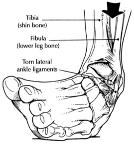
    Chronic ankle instability is the recurring “rolling” or “giving away” of the outer side of the ankle. The ligaments on the lateral side of the ankle may become weakened over time and after repeated ankle sprains. Typically the “rolling” or “giving away” occurs when walking or being active. This is a common occurrence with athletes. However, this can even happen when simply standing.

    Symptoms:

    Symptoms of chronic ankle instability may include pain, swelling or tenderness of the area. The most obvious symptom is the frequent rolling or turning of the ankle.

    Causes:

    Chronic ankle instability is generally caused by an ankle sprain that has not healed completely. When the ankle is sprained the ligaments are damaged. These ligaments, depending on the severity of the injury, are either stretched or torn. The image on the top right shows these ligaments as being wavy. This is how the ligaments appear after being stretched as a result of an ankle sprain. These stretched ligaments can lead to chronic ankle instability if they are not properly healed.

    Diagnosis:

    If you experience frequent ankle sprains or chronic instability in your ankles you should have this evaluated by us. If this condition is left untreated it can lead to further complications. The instability can lead to injuries as a result of a fall and arthritis and tendon problems. Depending on your circumstances an x-ray, CT scan, or MRI may be ordered to determine the best course of action.

    Treatment:

    Based on the examination results surgery may or may not be necessary. Nonsurgical treatments include physical therapy to rehabilitate and strengthen the muscles. Bracing can be effective by offering support to the ankle and prevent abnormal movement. Nonsteroidal anti-inflammatory drugs like ibuprofen can reduce swelling and pain. If the problem is severe surgery may be necessary. If the instability is significant and non-surgical methods do correct the issue reconstructive surgery to repair the ligaments will correct this issue.

    Other common causes of ankle pain

    There are many reasons for ankle pain; some of the most common include a history of ankle sprains or trauma.  Trauma or sprains can often result in inflammation of the ankle joint or damage to soft tissue structures like ankle ligaments or tendons. The anterior talar fibular ligament or peroneal tendons are often injured with sprains.

    Patients often relate a stiffness, popping or catching of the ankle as part of these symptoms.  These can be attributed to other causes including arthritis of the ankle or damage to cartilaginous surfaces. 


    There are a variety of treatment options from conservation care to surgical reconstruction. 

  • Foot and Ankle pain

    Quite a few patients, especially runners or athletes come in with:

    • ankle and foot pain
    • ankle and foot pain after running
    • ankle and foot pain without swelling

    There are many reasons for ankle pain, some of the most common include a history of ankle sprains or trauma.  Trauma or sprains can often result in inflammation of the ankle joint or damage to soft tissue structures like ankle ligaments or tendons.    The anterior talar fibular ligament or peroneal tendons are often injured with sprains.

     ankle knee pain

    Patiens often relate a stiffness, popping or catching of the ankle as part of there symptoms.  These can be attributed to other causes including arthritis of the ankle or damage to cartilaginous surfaces. 

    ankle conti anatomy01

    There are a variety of treatment options from conservation care to surgical reconstruction. 

    Common Foot And Ankle Disorders

    Did you know the foot has 26 bones, 33 joints, 107 ligaments, 19 muscles, and numerous tendons? These parts all work together to allow the foot to move in a variety of ways while balancing your weight and propelling you forward or backward on even or uneven surfaces. It is no wonder that 75 percent of all Americans will experience foot problems at one point or another in their lifetimes. At Issaquah Foot and Ankle Specialists we know what you're looking for in a foot doctor and we know we have the best foot care in the Seattle area. Our office can help diagnosis and treat your foot and ankle disorders from Flatfoot to Foot Reflexology, we consider all cases and all methods of pain relief. Click on the categories below to read articles on foot disorders and conditions.

    "What is a Podiatrist?"

    Most Common Disorders

    Foot Disorder Links

    Achilles Tendonitis

    Accessory Navicular Syndrome

    Adult Flatfoot

    Ankle Arthritis

    Ankle Fractures

    Ankle Pain

    Athlete's Foot

    Bunion Deformity

    Calcaneal Apophysitis

    Capsulitis of the Second Toe

    Charcot Foot

    Chronic Ankle Instability

    Fungal Toenails

    Gout

    Hallux Limitus

    Hallux Rigidus

    Hammertoes

    Heel Pain: Heel Spurs

    Heel Pain: Plantar Fasciitis

    Ingrown Toenails

    Metatarsal Stress Fracture

    Neuroma

    Plantar Fasciitis: Arch Pain

    Posterior Tibial Tendon Dysfunction (PTTD)

    Posterior Tibial Tendonitis

    Sesamoids

    Shin Splints

    Tailor's Bunion 

    Achilles Tendon Problems

    Ankle Injuries

    Ankle Surgery

    Bone Tumors

    Circulation Disorders

    Diabetic Patient Information

    Diseases of the Skin

    Diseases of the Toenails

    Fitting Shoes

    Foot Surgery

    General Disorders

    Knee Problems In Children

    Lumps and Bumps

    Nerve Conditions

    Pain Experienced by the Walking Child

    Pain in the Foot by Location

    Pediatric and Childhood Abnormalities

    Soft Tissue Masses

    Special Testing Equipment

    Sports Injuries

    Tendon Injuries

    Toe Problems

    Trauma and Injury to the Foot

    Treatments


  • Peroneal Tendon Dysfunction
    Dislocating peroneal tendons are an uncommon injury to a group of two tendons whose muscles originate on the outside of the calves. These two muscles are named the Peroneus Brevis and Peroneus Longus. These two muscles are responsible for eversion of the foot. This movement of the foot is demonstrated by standing and then rolling to the outside of the foot. These tendons are also called "stirrup" tendons because as they pass into the foot they act as a stirrup to help hold up the arch of the foot. As these tendons pass behind the outside ankle bone, called the fibula, they are held in place by a band of tissue called the peroneal retinaculum. Injury to the retinaculum can cause it to stretch or even tear. When this occurs the peroneal tendons can dislocate from their groove on the back of the fibula. The tendons can be seen to roll over the outside of the fibula. This will cause the tendons to function abnormally and can cause damage to the tendons. Dislocating peroneal tendons most commonly occur as a result of injury during participation in athletic activities. The most common sport causing injury is snow skiing. It can also occur while playing football, basketball, and soccer. This injury can occur in non-athletes, as a result of a severe ankle sprain. The injury typically results in a popping or sharp pain on the outside of the ankle. The outside of the ankle is called the lateral malleolus. Commonly however, there may be little to no discomfort at the time of injury, which later becomes symptomatic.

    Diagnosis

    Physical examination will reveal swelling behind the outside of the ankle if it is an acute injury. If the injury is chronic there may be little to no swelling. There is usually tenderness particularly when pressure is applied behind the outside of the ankle. Having the patient forcefully turn the foot outward against the physician's hand can demonstrate dislocation of the peroneal tendons. This will cause the peroneal tendons to dislocate over the outer edge of the lateral malleolus.

    X-rays and occasionally an MRI exam may be necessary to confirm the diagnosis. X-rays are commonly taken to ensure that there are not any other injuries to the bones of the foot and ankle. An MRI will provide your physician with information about abnormal positioning and/or possible tears of the peroneal tendons.

    Treatment

    Non Surgical Treatment

    If there is an acute injury the initial treatment is usually crutches with no weight being applied to the foot. Usually a splint or compressive wrap is applied to decrease swelling. Anti-inflammatory medications and ice are often utilized to help decrease swelling. Once the swelling has subsided your physician will be better able to tell the true extent of injury. Mild injury can be treated with conservative, non-surgical means. Mild injury results in stretching of the peroneal retinaculum without dislocation of the peroneal tendons. The patient is placed in a cast or removable cast boot and must use crutches for six weeks. During this time no weight can be applied to the foot. After six weeks the patient is re-examined. If the injury has not healed further treatment may be necessary.

    Surgical Treatment

    Surgical correction is necessary in cases of failed conservative therapy and moderate to severe injuries. With moderate to severe injuries the peroneal retinaculum is either torn or severely stretched to a point that the peroneal tendons will easily dislocate. Surgery will involve tightening the stretched or torn peroneal retinaculum. Both absorbable and non-absorbable suture or stitches will be necessary to hold the tissue in place until it heals. This may require drill holes or metallic anchors to be placed in the fibula to aid in suturing the peroneal retinaculum back onto itself.

    Twenty-five percent of the population does not have a groove on the back of the fibula for the peroneal tendons to move in. This groove is called the peroneal groove. This can be a causative factor for peroneal tendon dislocation and can only be identified at the time of surgery. If this is encountered during surgery a peroneal groove will be created by performing bone cuts in the back of the fibula. These may require bone screws or pins to hold the bone in place while it heals. During the surgery the peroneal tendons will be examined for possible tears or damage. If this is found it will be repaired by suture.

    Post-operatively the patient is placed on crutches and in a splint or cast for 4 to 6 weeks with no weight being applied to the foot. This is followed by 2 to 4 weeks of protected weight bearing in a cast or removable cast boot. This is usually followed by 3 to 6 weeks of physical therapy to regain strength and motion.

    Potential Complications

    Once an injury occurs it is always possible for a person to re-injure or re-dislocate their peroneal tendons. Surgical complications are rare but can include: infection, reoccurrence, stiffness and weakness of the peroneal tendons, and delay or failure of bone cuts in the fibula to heal. The risk for complications is greatly reduced by taking your prescriptions as instructed and strictly following post-operative instructions.

  • Ankle Fusion/Arthrodesis
    What is an ankle arthrodesis?

    The ankle joint consists of portions of three bones: the talar dome, distal tibial plafond and distal fibula. The talus and tibia portions make up over 80% of the articular surface area of the ankle joint. Arthrodesis procedures are the removal of cartilage and any diseased bone from a joint to produce a fusion of at least two bones to create one bone. This removal of cartilage exposes the underlying bone on both sides of the joint. These joints surfaces are then compressed together with some form of fixation to create the fusion. In an ankle arthrodesis, typically the tibial position is fused to the talus. In some cases the fibula is part of arthrodesis, but this is the surgeon’s preference.

    Why is an ankle arthrodesis procedure performed?

    Ankle arthrodesis procedures are typically performed when all conservative options have been attempted and failed. Some of these treatment options, are corticosteroid injections, ankle foot orthoses, anti-inflammatory medications, custom orthotics, ankle braces, and sometimes arthrotomy or arthroscopy. In cases where the pain and/or the deformity is unremitting the arthrodesis is recommended. Some of the conditions that produce unremitting pain and deformity in the ankle joint are:

    1. Post-traumatic arthritis
    2. Rheumatoid arthritis
    3. Infection
    4. Failed ankle implant
    5. Congenital ankle deformities
    6. Neuromuscular disorders (i.e. post-polio syndrome, paraplegia, etc.)
    7. Lateral ankle instability (after failure of operative and non-operative treatment)
    8. Avascular Necrosis (secondary to trauma or metabolic disease states)
    9. Charcot Neuropathy

    Techniques for ankle arthrodesis

    Over the years many techniques have been devised to fuse the ankle joint. Today there are three different techniques that your surgeon may use. They are open technique (use of one or two incisions on the outside and the front of your ankle) with screw fixation, open technique with external fixator fixation and arthroscopic ankle arthrodesis. Most surgeons use all three of these techniques, with the open technique with screw fixation the most commonly used. The technique used is surgeon's preference for your clinical findings, but all have equal fusion rates. Sometimes your doctor may need the use of bone graft, typically taken from your hip area (iliac crest), if you have defects in you ankle from previous trauma or surgery to help support the arthrodesis site.

    What to Expect Following the Procedure

    After most ankle arthrodesis procedures, your surgeon will have you non-weightbearing (no weight on your foot) typically for a period of 6-12 weeks, but this can be extended longer if there is any delay in healing of the arthrodesis site. After the surgery, you will be in a below knee cast or splint to help protect the surgical site and also prevent any movement. After the 6-12 weeks period your surgeon, will typically start you partially weightbearing with your crutches with the use of some type of cast or ankle brace for 4-6 weeks. Once you are fully weightbearing in the cast brace, you will be progressed into high-topped shoe or sneaker. Sometimes modifications have to be made to your shoes, called a rocker-bottomed sole, to aid in push off in ambulation after surgery.

    Some other things that are important to note after the surgery, typically there will be some level of pain or discomfort after the surgery. In a majority of cases, you will be admitted after the surgery to the hospital for pain management. This admission can range from 23 hour to 3-4 days based on an individual’s pain tolerance. When in the hospital and also when you go home it is essential that you keep your foot elevated and use ice as recommended by your surgeon. Ice and elevation will help to reduce the swelling around your foot and ankle that is common after a major surgical reconstruction. The swelling in your foot and ankle can last for 6-9 months and even up to a year. Another thing that is tied closely to the swelling after surgery is a phenomenon that occurs when you get up from after you have had your foot elevated for a period of time. What happens is when you dangle your leg to get up, blood flow will increase into the foot and ankle which will create a throbbing sensation and sometimes a pins and needles sensation in your foot. This is perfectly normal and will go away when you re-elevate your foot.

    Recovery Time

    Time off work depends on the type of ones work as well as the procedures performed. Usually a patient can return to work in 2 to 4 weeks if they are able to work while seated. If a persons job requires standing and walking, return to work may take several weeks when one is able to bear weight. Complete recovery may take six months to a full year.

    Potential Complications

    Complications can occur as with all surgeries, but are minimized by strictly following your surgeons post-operative instructions. The main complications include infection, bone that is slow to heal or does not heal, progression or reoccurrence of deformity, a stiff foot, and the need for further surgery. Many of the above complications can be avoided by only putting weight on the operative foot when allowed by your surgeon.

  • Ankle Arthritis

    Ankle arthritis can result from such things as ankle sprains, ankle fractures and infection.  Other conditions that can lead to an arthritic ankle include rheumatoid arthritis or other inflammatory conditions. 

    Ankle arthritis treatment in the Issaquah, WA 98027 area

    Ankle Arthritis results from  a loss of cartilage within the joint that normally provides a lubricated surface for smooth range of motion.  The cartilage begins to wear away and patients often feel a catching, stiffness, swelling or pain with activity.

    Treatment for ankle arthritis can include conservative or surgical care.

    Conservative care:

    • Bracing
    • Injections
    • NSAIDS
    • Physical Therapy

    Surgical care:

    Ankle Arthroscopy in the Issaquah, WA 98027 area

    • Ankle Arthroscopy
    • Ankle Arthrotomy
    • Ankle Arthrodesis
    • Ankle Joint Replacement
    • Ankle Arthrodiatasis
5 out of 5 stars
Total Reviews : 232