vendor

vendor

vendor

Friday, 05 April 2019 13:35

Insanity and Achilles Tendonitis!


Achilles tendinitis can be one of the more stubborn conditions that we treat. We have come up with very effective protocols to treat this. We do like to follow-up and make certain that our patients are getting better. And I like to check that the symptoms are progressively improving and in most cases completely resolving. There are several different "layers" of treatment. There is what I consider the core treatments. Most of these treatments are mechanical in nature as is the case with many foot and ankle problems. But some of the adjunctive treatments also address the underlying inflammatory or in some cases degenerative processes that can occur.


The core treatments: special prescription orthotics, special Achilles exercises, a night splint, and a special Achilles daytime and exercise brace. Note, many clinics do not know the most effective orthotics or the best braces for this condition. The secondary additional options include: shockwave treatment, cortisone injections, PRP (platelet rich plasma), gastroc recession procedure, and Achilles surgical procedures. Again, many clinics do not have experience with these advanced treatment options.


It is important to include all of the core treatments to help this problem resolve and keep it from coming back. Once the core treatments have been provided, if there are still symptoms then there is the option of the adjunctive treatments which can be especially helpful for the stubborn cases. We provide all of these treatments at our clinic. Some of our patients come in and have had only minor improvement with their Achilles problems. They come to our clinic  for these other options. However, it always surprises me when a new patient comes in and they do not want any of the newer treatment options. They only want to do the same treatments that were done before at other clinics that did not work. And, surprise they still have their Achilles problems!


What do they say? “Insanity is doing the same thing over and over again and expecting different results"!


We offer all of these treatments that I mention at our clinic. The treatments that are not being provided may be holding up your progress! Give us a call at 425-391-8666 or make an appointment online today to see a foot and ankle specialist.

One of the most exciting new technologies available for shin splints is platelet rich plasma therapy. Shin splints can be a large source of frustration for avid exercisers or runners. We see quite a few patients that have had increased pain and discomfort daily with running and have tried multiple treatment modalities all to no avail. Patients have often tried changing shoes, physical therapy, stretching, icing and anti-inflammatories and have had to stop running or change training techniques to try to help with their shin splints.

Research has shown for decades that shin splints are caused by inflammation. Treating the root cause of the inflammation with platelet rich plasma therapy has shown extremely promising results. Platelet rich plasma therapy involves using your own body's naturally healing enzymes at the site of an inflammatory pain to stop the process altogether. The procedure itself can be performed in the office. We're excited to be able offer this to patients who have suffered from shin splints and want to return to activities. If you suffer from shin splints we can fix it. Give our office a call today at 425-391-8666 or make an appointment online.

Friday, 29 March 2019 03:27

Equinus Part 2

How does a large calf muscle mimic a tight Achilles and equinus? Some individuals just have a relatively large calf muscle. All that extra muscle mass can exert a disproportionate mechanical pull on the Achilles tendon and ultimately during gait this scenario can have a similar effect to a tight calf muscle (equinus). The constant pulling on the foot can lead to the same problems that an individual with a tight calf muscle or gastroc equinus has.

Those individuals are prone to plantar fasciitis, and forefoot problems. Stretching the calf muscle is always a good idea and especially with these individuals. If an individual is unlucky enough to have both a large strong calf muscle and a tight calf muscle it really compounds the problems for foot biomechanics. These individuals are extremely prone to problems including plantar fasciitis and forefoot problems. There are also prone to mid foot problems. If you are experiencing foot or ankle pain, give us a call at 425-391-8666 or make an appointment online. 

Friday, 22 March 2019 02:08

Equinus Part 1

Basically equinus means a tight calf (or tight Achilles) and the tendency to have limited ankle joint range of motion.This can be similar to a person wearing high-heeled shoes and the heel is off the ground or quickly elevates off the ground during gait and weight is rapidly transferred to the ball the foot.This also means there is limited ability of the ankle and its ability to flex upward with the top of the foot toward the front of the leg.The ball of the foot which would include the weightbearing metatarsals in the forefoot, receive much more stress load than they are designed to take.The load is increased faster than normal and during a normal gait cycle they receive a disproportionate amount of weightbearing time and force.

So if there is any slight imbalance on the ball the foot, the equinus force will result in increased stress load in the wrong place.A very similar condition can result when patient presents with large muscular calves and all that muscle bulk has the tendency to pull the heel up quickly.Some individuals and especially some children are "toe walkers". Either they walk around on the ball of her feet or as they are walking that heel comes up quite quickly or they have a "bouncy" gait were the heel just rapidly lifts up.There can be a gastrocnemius equinus or shortening of the gastroc muscle or a combination of gastrocnemius and soleus muscle equinus.Gastrocnemius equinus seems to be the most common.

If you are experience foot or heel pain, give us a call at 425-391-8666 or make an appointment online today.

Friday, 15 March 2019 12:07

Bone Healing After Surgery

After surgery, there is bone healing involved, such as a midfoot fusion or resetting of the bone – and osteotomy. The big question is when has that bone healed enough that you can put full weight on it without a regular cast or even a cast boot. We always check this with x-rays. X-rays are helpful but seem to lag behind what’s actually going on deep inside. This lag can be several weeks or longer.


So I often use ultrasound to look inside. The ultrasound image will go to the soft tissue and stop at the bone. But if there’s a small gap at the plant fusion site it will show this. If there’s some areas of bridging it will also show that. I will look around the fusion site or osteotomy site circumferentially and if I see areas of definitive bridging that is a very good sign that often is weeks ahead of the x-ray image.

Friday, 15 March 2019 12:05

Cartiva Implant for Big Toe Arthritis

One of the latest procedures for arthritis of the big toe involves the use of an implant to eliminate pain and maintain motion. Traditionally, we have elected to fuse or arthrodesis the big toe. However this gold standard is shifting. A lot of patients want to maintain motion or movement of the big toe for activities like yoga. We are seeing quite a few patients elect for an implant of the big toe instead of the fusion.

The recovery is much quicker, the procedure takes less time and is less invasive. We are seeing promising results with the Cartiva Implant and are excited to be offering this at our clinic. With our own surgery center we can provide a significant time and cost savings. Instead of patients staying at the hospital for half a day we can usually get patients in and out in hours. The Cartiva is a great advancement in the care of the arthritic joint and we are excited to see where it take our patients. Give us a call at 425-391-8666 or make an appointment online today.

Friday, 08 March 2019 12:04

Removal of Screws in the Foot and Ankle

After some types of foot or ankle surgery there will be hardware used such as screws or plates. When removing the screws it can be helpful to plan out the exact location of the head of the screw so that the skin incision can be smaller and more precisely located. Therefore there often is faster healing with a smaller wound (and scar) and less postoperative symptoms or pain.


So I often use ultrasound imaging prior to the surgery and map out the location of the screw head. I also sometimes will use fluoroscopy and place a metallic reference-object over the skin when I do the floroscopic image. Again with either one I can plan ahead prior to surgery for shorter surgical time itself and a less involved procedure.

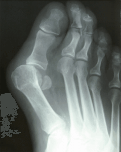
Bunion xray


Can you have both the bunion and a ganglion cyst in the same location?


The answer is yes. A ganglion cyst is a cyst that is filled with a viscous clear fluid. This fluid usually comes from an underlying joint or tendon sheath. With a traditional bunion, the great toe starts deviating laterally, and therefore the medial capsule becomes thinner. This thinning capsule is also weaker and it's easier for the inner fluid to herniate out starting the ganglion cyst structure. On x-ray there will be a bunion but increased soft tissue density or thickness of the soft tissue structures over the bunion itself. This ganglion cyst can also be diagnosed with ultrasound imaging. In some cases, the cyst will be soft and compressible on top of the bunion and will seem spongy to exam or palpation. 


The ganglion cyst superimposed over the bunion will also make the bunion look worse than the bunion really is structurally. Therefore with an x-ray, one will appreciate the bunion just does not look as bad as you expect compared to when you look at your foot. If you have a bunion or a cyst-like growth on your foot and would like further evaluation please let us know. Either myself Dr. Timothy Young, or my partner Dr. Brandon Nelson would be happy to evaluate this. We have on-site x-rays and ultrasound imaging if necessary. Give us a call at 425-391-8666 or make an appointment online today.

Friday, 01 March 2019 12:00

How to Fix Hammertoes

Hammertoes are a common pathology of the lower extremity. Quite a few people have a bunion associated with hammertoes and this makes it a little more challenging to fix. The hammertoe can be created from a much different pathology. The most common pathology, however, is from a compensatory mechanism to stabilize the foot structure. The most common being the flexor tendons on the bottom of the foot firing to help control somebody with flatfoot for an unstable arch.

Hammertoes can be broken down and into different types, one being a flexible hammertoe deformity and the second being a fixed hammertoe deformity. Flexible hammertoe deformities are often amenable to conservative measures like calf stretching and bracing. However the majority of fixed hammertoes require surgical intervention. There are many different types of procedures that can be done for hammertoes and the majorities are done in outpatient procedure setting and can be performed in less than 30 minutes. Hammertoes can continue to progress so early intervention is key to slowing down the deformity itself. Make an appointment to see a foot and ankle specialist today or give us a call at 425-391-8666.


Stress fractures in the foot and metatarsals (forefoot) are quite common. There’s almost always swelling of the forefoot and often there is pain. It is most common with the second metatarsal but it can also involve the third or fourth metatarsals. The first and fifth and metatarsals are much less common.


With a stress fracture there is bone fatigue to the point, that there’s a break in the outer shell or cortex of the bone. But it’s a hairline break in the body usually is able to try to protect it and start the process of healing. The body‘s way to heal this is to form a bone callous adjacent to the break in the bone. The bone callous becomes is visible on x-ray where as the original fracture is such a fine hairline break that often you cannot see it on x-ray. With enough time or activity the fracture outer shell cortical fracture or break can progress to a displaced fracture that is now a visible fracture on x-ray. 


On the other hand, with a stress reaction of bone there is not a break in the cortex of the bone and the bone callous therefore never develops. But inside the bone there is still marrow edema. This means that the bone has increased activity and literally swelling within the bone. With both a stress reaction of bone and a stress fracture there is marrow edema that can be seen with an MRI - for early detection of both conditions. Ultrasound imaging can often show a stress fracture early, but not a stress reaction of bone.


Functionally both conditions are quite similar since they both involve bone healing. Typical bone healing for an adult is eight weeks. If you have pain in your feet or swelling, please make an appointment for a consultation at our clinic or give us a call at 425-391-8666.

Page 2 of 26
5 out of 5 stars
Total Reviews : 232